1 Department of Molecular Pharmacology and Experimental Therapeutics, Mayo Clinic, Rochester, Minnesota.
2 Center for Regenerative Medicine, Mayo Clinic, Rochester, Minnesota.
Stem Cells Dev. 2019 May 15;28(10):659-673. doi: 10.1089/scd.2019.0030. Epub 2019 Apr 17.
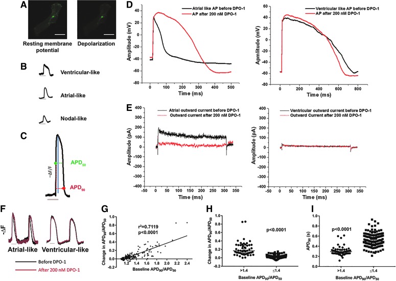
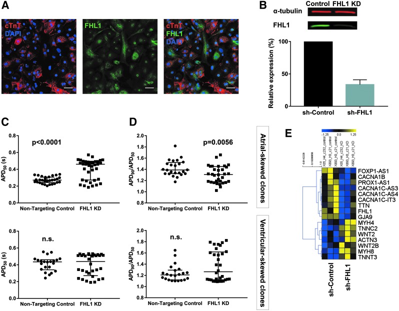
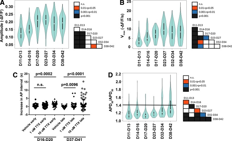
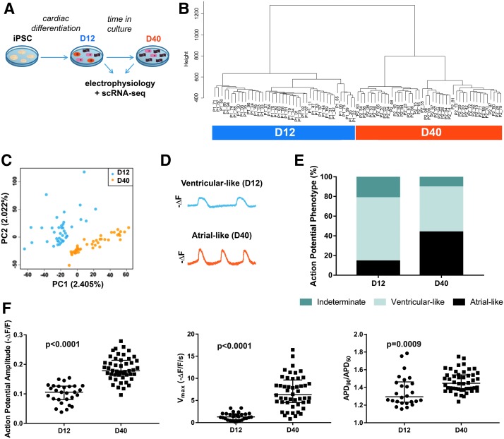
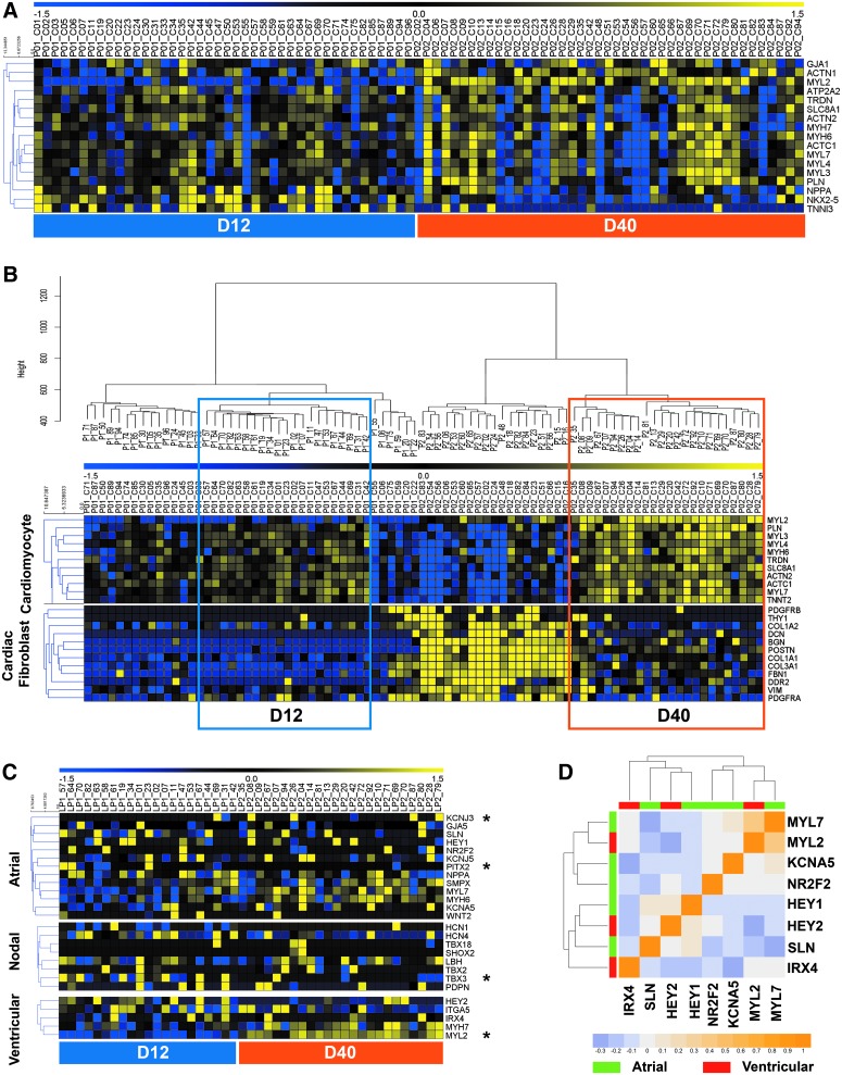
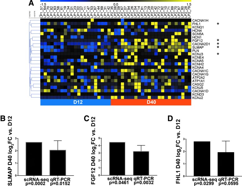
The ability to accurately phenotype cells differentiated from human induced pluripotent stem cells (hiPSCs) is essential for their application in modeling developmental and disease processes, yet also poses a particular challenge without the context of anatomical location. Our specific objective was to determine if single-cell gene expression was sufficient to predict the electrophysiology of iPSC-derived cardiac lineages, to evaluate the concordance between molecular and functional surrogate markers. To this end, we used the genetically encoded voltage indicator ArcLight to profile hundreds of hiPSC-derived cardiomyocytes (hiPSC-CMs), thus identifying patterns of electrophysiological maturation and increased prevalence of cells with atrial-like action potentials (APs) between days 11 and 42 of differentiation. To profile expression patterns of cardiomyocyte subtype-associated genes, single-cell RNA-seq was performed at days 12 and 40 after the populations were fully characterized with the high-throughput ArcLight platform. Although we could detect global gene expression changes supporting progressive differentiation, individual cellular expression patterns alone were not able to delineate the individual cardiomyocytes into atrial, ventricular, or nodal subtypes as functionally documented by electrophysiology measurements. Furthermore, our efforts to understand the distinct electrophysiological properties associated with day 12 versus day 40 hiPSC-CMs revealed that ion channel regulators , , and were the most significantly increased genes at day 40, categorized by electrophysiology-related gene functions. Notably, knockdown during differentiation was sufficient to significantly modulate APs toward ventricular-like electrophysiology. Thus, our results establish the inability of subtype-associated gene expression patterns to specifically categorize hiPSC-derived cells according to their functional electrophysiology, and yet, altered expression is able to redirect electrophysiological maturation of these developing cells. Therefore, noncanonical gene expression patterns of cardiac maturation may be sufficient to direct functional maturation of cardiomyocytes, with canonical gene expression patterns being insufficient to temporally define cardiac subtypes of in vitro differentiation.
准确表型分化自人类诱导多能干细胞(hiPSCs)的细胞对于将其应用于发育和疾病过程的建模至关重要,但在没有解剖位置背景的情况下,这也是一个特别的挑战。我们的具体目标是确定单细胞基因表达是否足以预测 iPSC 衍生的心脏谱系的电生理学,评估分子和功能替代标志物之间的一致性。为此,我们使用遗传编码的电压指示剂 ArcLight 对数百个 hiPSC 衍生的心肌细胞(hiPSC-CMs)进行了分析,从而确定了电生理成熟的模式和分化第 11 天至第 42 天之间具有类似心房动作电位(AP)的细胞的发生率增加。为了分析与心肌细胞亚型相关的基因的表达模式,在高通量 ArcLight 平台对细胞群体进行充分表征的第 12 天和第 40 天后进行了单细胞 RNA-seq。尽管我们可以检测到支持逐渐分化的全局基因表达变化,但单独的细胞表达模式本身并不能将单个心肌细胞划分为心房、心室或结状亚型,正如电生理学测量所记录的那样。此外,我们努力了解与第 12 天和第 40 天 hiPSC-CMs 相关的独特电生理特性,结果表明,离子通道调节剂 、 和 是第 40 天表达最显著增加的基因,根据与电生理学相关的基因功能进行分类。值得注意的是,在分化过程中 的敲低足以显著将 AP 调节为类似心室的电生理学。因此,我们的结果表明,与亚型相关的基因表达模式无法根据其功能电生理学将 hiPSC 衍生的细胞专门分类,但是改变 的表达能够重新定向这些发育中的细胞的电生理成熟。因此,心脏成熟的非典型基因表达模式可能足以指导心肌细胞的功能成熟,而典型的基因表达模式不足以在时间上定义体外分化的心脏亚型。