Department of Biomedical Imaging and Image-Guided Therapy, Division of Cardiovascular and Interventional Radiology, Medical University of Vienna, Vienna, Austria.
Department of Biomedical Imaging and Image-Guided Therapy, Division of Nuclear Medicine, Medical University of Vienna, Vienna, Austria.
J Magn Reson Imaging. 2019 Oct;50(4):1326-1335. doi: 10.1002/jmri.26722. Epub 2019 Mar 20.
Sympathetic reinnervation after heart transplantation (HTX) is a known phenomenon, which has an impact on patient heart rate variability and exercise capacity. The impact of reinnervation on myocardial structure has not been evaluated yet.
To evaluate the feasibility of simultaneous imaging of cardiac reinnervation and cardiac structure using a hybrid PET/MRI system.
Prospective / pilot study.
Ten patients, 4-21 years after cardiac transplantation.
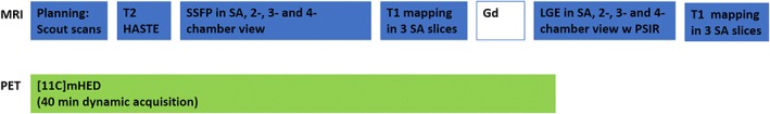
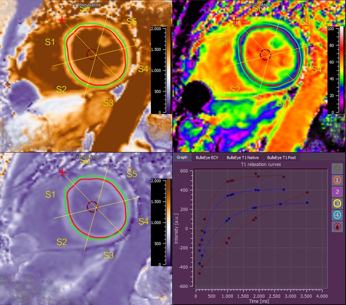
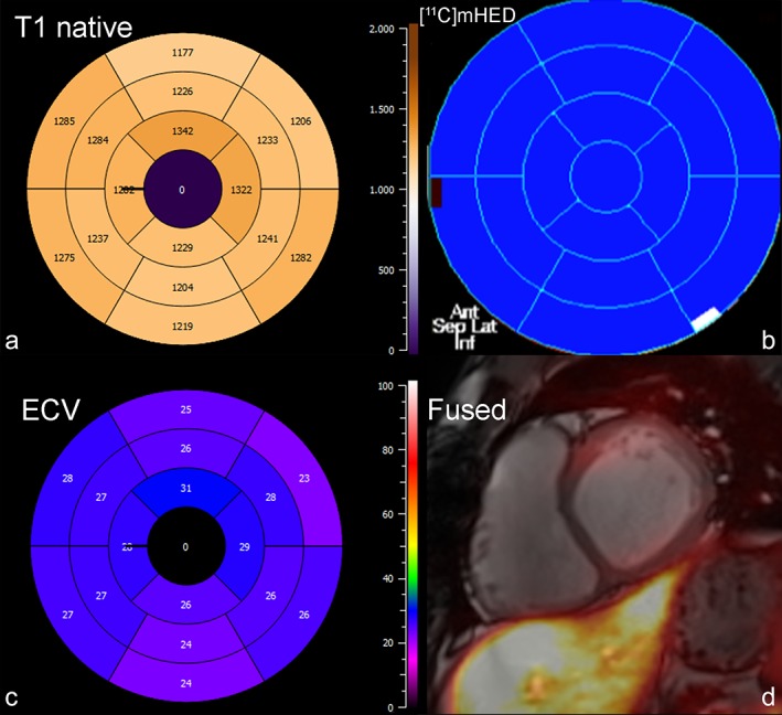
FIELD STRENGTH/SEQUENCE: 3 T hybrid PET/MRI system. Cine SSFP, T mapping (modified Look-Locker inversion recovery sequence) pre/postcontrast as well as dynamic [ C]meta-hydroxyephedrine ([ C]mHED) PET.
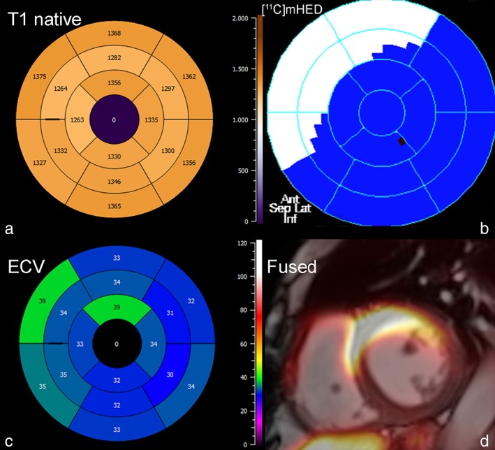
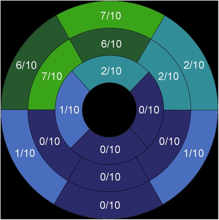
All MRI and PET parameters were evaluated by experienced readers using dedicated postprocessing software packages for cardiac MRI and PET. For all parameters a 16-segment model for the left ventricle was applied.
Mann-Whitney U-test; Spearman correlations.
Thirty-six of 160 myocardial segments showed evidence of reinnervation by PET. On a segment-based analysis, mean native T relaxation times were nonsignificantly altered in segments with evidence of reinnervation (1305 ± 151 msec vs. 1270 ± 112 msec; P = 0.1), whereas mean extracellular volume (ECV) was significantly higher in segments with evidence of reinnervation (35.8 ± 11% vs. 30.9 ± 7%; P = 0.019). There were no significant differences in wall motion (WM) and wall thickening (WT) between segments with or without reinnervation (mean WM: 7.6 ± 4 mm vs. group B: 9.3 ± 7 mm [P = 0.13]; WT: 79 ± 63% vs. 94 ± 74% [P = 0.27]) under resting conditions.
The assessment of cardiac reinnervation using a hybrid PET/MRI system is feasible. Segments with evidence of reinnervation by PET showed nonsignificantly higher T relaxation times and a significantly higher ECV, suggesting a higher percentage of diffuse fibrosis in these segments, without impairment of rest WM and WT.
3 Technical Efficacy: Stage 3 J. Magn. Reson. Imaging 2019;50:1326-1335.
心脏移植(HTX)后交感神经再支配是一种已知的现象,它会影响患者的心率变异性和运动能力。再支配对心肌结构的影响尚未得到评估。
评估使用混合 PET/MRI 系统同时成像心脏再支配和心脏结构的可行性。
前瞻性/初步研究。
10 名心脏移植后 4-21 年的患者。
磁场强度/序列:3T 混合 PET/MRI 系统。电影 SSFP、T 映射(改良 Look-Locker 反转恢复序列)增强前后以及动态 [C]meta-羟基苯丙胺([C]mHED)PET。
所有 MRI 和 PET 参数均由经验丰富的读者使用心脏 MRI 和 PET 的专用后处理软件包进行评估。对于所有参数,均应用左心室 16 节段模型。
曼-惠特尼 U 检验;Spearman 相关。
160 个心肌节段中有 36 个通过 PET 显示出再支配的证据。基于节段分析,在有再支配证据的节段中,原生 T 弛豫时间的平均值无明显变化(1305±151msec 与 1270±112msec;P=0.1),而有再支配证据的节段的细胞外容积(ECV)平均值明显较高(35.8±11%与 30.9±7%;P=0.019)。有再支配和无再支配节段之间的壁运动(WM)和壁增厚(WT)没有显著差异(平均 WM:7.6±4mm 与 B 组:9.3±7mm [P=0.13];WT:79±63%与 94±74% [P=0.27])。
使用混合 PET/MRI 系统评估心脏再支配是可行的。通过 PET 显示出再支配证据的节段显示出 T 弛豫时间升高不明显,而 ECV 升高显著,提示这些节段中弥漫性纤维化的百分比更高,但不影响静息 WM 和 WT。
3 级技术功效:第 3 阶段 J. Magn. Reson. Imaging 2019;50:1326-1335。