• 文献检索
  • 文档翻译
  • 深度研究
  • 学术资讯
  • Suppr Zotero 插件Zotero 插件
  • 邀请有礼
  • 套餐&价格
  • 历史记录
应用&插件
Suppr Zotero 插件Zotero 插件浏览器插件Mac 客户端Windows 客户端微信小程序
定价
高级版会员购买积分包购买API积分包
服务
文献检索文档翻译深度研究API 文档MCP 服务
关于我们
关于 Suppr公司介绍联系我们用户协议隐私条款
关注我们

Suppr 超能文献

核心技术专利:CN118964589B侵权必究
粤ICP备2023148730 号-1Suppr @ 2026

文献检索

告别复杂PubMed语法,用中文像聊天一样搜索,搜遍4000万医学文献。AI智能推荐,让科研检索更轻松。

立即免费搜索

文件翻译

保留排版,准确专业,支持PDF/Word/PPT等文件格式,支持 12+语言互译。

免费翻译文档

深度研究

AI帮你快速写综述,25分钟生成高质量综述,智能提取关键信息,辅助科研写作。

立即免费体验

定量心血管磁共振:再血管化心肌梗死后患者的细胞外容积、原生 T1 和 18F-FDG PET/CMR 成像及其与心肌损伤和全身炎症标志物的相关性。

Quantitative cardiovascular magnetic resonance: extracellular volume, native T1 and 18F-FDG PET/CMR imaging in patients after revascularized myocardial infarction and association with markers of myocardial damage and systemic inflammation.

机构信息

Nuklearmedizinische Klinik und Poliklinik, Klinikum rechts der Isar, Technische Universität München, Ismaninger Straße 22, 81675, Munich, Germany.

Klinik und Poliklinik für Medizin I, Klinikum rechts der Isar, Technische Universität München, Ismaninger Straße 22, 81675, Munich, Germany.

出版信息

J Cardiovasc Magn Reson. 2018 May 24;20(1):33. doi: 10.1186/s12968-018-0454-y.

DOI:10.1186/s12968-018-0454-y
PMID:29792210
原文链接:https://pmc.ncbi.nlm.nih.gov/articles/PMC5967072/
Abstract

BACKGROUND

Characterization of tissue integrity and inflammatory processes after acute myocardial infarction (AMI) using non-invasive imaging is predictive of patient outcome. Quantitative cardiovascular magnetic resonance (CMR) techniques such as native T and extracellular volume (ECV) mapping as well as F-FDG positron emission tomography (PET) imaging targeting inflammatory cell populations are gaining acceptance, but are often applied without assessing their quantitative potential. Using simultaneously acquired PET/CMR data from patients early after AMI, this study quantitatively compares these three imaging markers and investigates links to blood markers of myocardial injury and systemic inflammatory activity.

METHODS

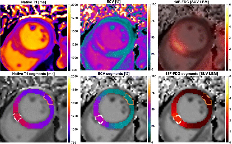
A total of 25 patients without microvascular obstruction were retrospectively recruited. All imaging was simultaneously performed 5 ± 1 days after revascularization following AMI on an integrated 3T PET/MRI scanner. Native and post-contrast T data were acquired using a modified Look-Locker inversion recovery (MOLLI) sequence, ECV maps were calculated using individually sampled hematocrit. F-FDG PET was executed after 1 day of dietary preparation, 12 h of fasting, and administration of heparin. ECV, F-FDG and native T data were compared mutually as well as to peak counts of peripheral blood markers (creatine kinase, creatine kinase-MB, troponin, leukocytes, monocytes) and infarct size.

RESULTS

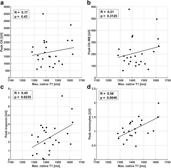
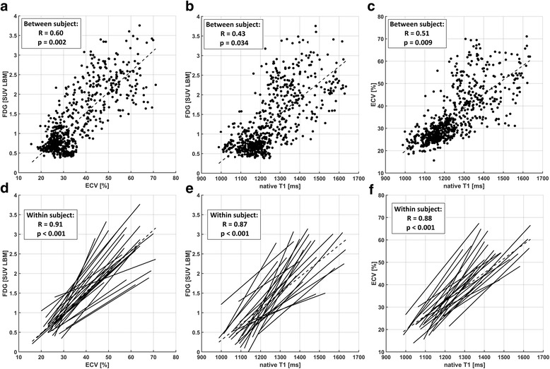
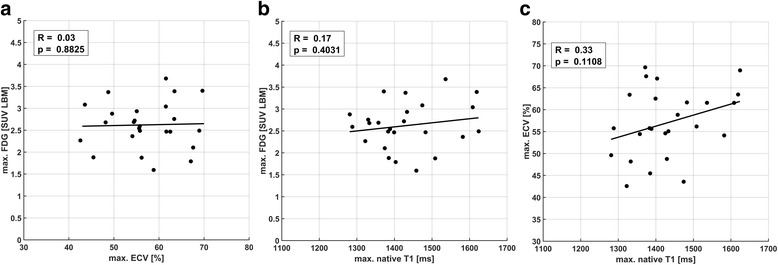
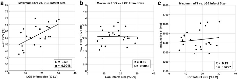
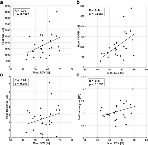
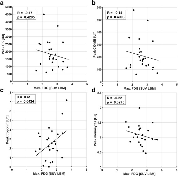
High intra-patient correlations of relative ECV, F-FDG PET and native T signal increases were observed in combination with no inter-patient correlation of maximum absolute values at the infarct center, suggesting well-colocalized but physiologically diverse processes begetting the respective image signals. Comparison of maximum image signals to markers of myocardial damage and systemic inflammation yielded highly significant correlations of ECV to peak creatine kinase-MB and overall infarct size as well as between native T and peak monocyte counts.

CONCLUSIONS

Absolute native T values at the infarct core early after AMI can be linked to the systemic inflammatory response independent of infarct size. Absolute ECV at the infarct core is related to both infarct size and blood markers of myocardial damage.

摘要

背景

使用非侵入性成像技术对急性心肌梗死(AMI)后组织完整性和炎症过程进行特征描述可预测患者的预后。定量心血管磁共振(CMR)技术,如天然 T 和细胞外容积(ECV)映射以及针对炎症细胞群的 F-FDG 正电子发射断层扫描(PET)成像,正逐渐得到认可,但在应用时通常并未评估其定量潜力。本研究使用 AMI 后早期同时采集的 PET/CMR 数据,定量比较了这三种成像标志物,并研究了它们与心肌损伤和全身炎症活性的血液标志物之间的联系。

方法

共回顾性招募了 25 名无微血管阻塞的患者。所有成像均在 AMI 后血管重建后 5±1 天在集成的 3T PET/MRI 扫描仪上同时进行。使用改良 Look-Locker 反转恢复(MOLLI)序列采集天然和对比后 T 数据,使用个体采集的红细胞压积计算 ECV 图。F-FDG PET 在进行 1 天的饮食准备、12 小时禁食和肝素给药后执行。ECV、F-FDG 和天然 T 数据相互比较,也与外周血标志物(肌酸激酶、肌酸激酶同工酶-MB、肌钙蛋白、白细胞、单核细胞)和梗死面积的峰值计数进行比较。

结果

在组合中观察到相对 ECV、F-FDG PET 和天然 T 信号增加的患者内高相关性,而在梗死中心的最大绝对值没有患者间相关性,这表明存在具有不同生理特征的高度聚集但不同的过程,从而产生各自的图像信号。将最大图像信号与心肌损伤和全身炎症标志物进行比较,发现 ECV 与肌酸激酶同工酶-MB 的峰值和整体梗死面积以及天然 T 与单核细胞峰值计数之间具有高度显著的相关性。

结论

AMI 后早期梗死核心处的绝对天然 T 值可与梗死大小无关的全身炎症反应相关联。梗死核心处的绝对 ECV 与梗死面积和心肌损伤的血液标志物均相关。

https://cdn.ncbi.nlm.nih.gov/pmc/blobs/21c7/5967072/4b46a43beeed/12968_2018_454_Fig7_HTML.jpg
https://cdn.ncbi.nlm.nih.gov/pmc/blobs/21c7/5967072/ca75b1868525/12968_2018_454_Fig1_HTML.jpg
https://cdn.ncbi.nlm.nih.gov/pmc/blobs/21c7/5967072/78b369f3a512/12968_2018_454_Fig2_HTML.jpg
https://cdn.ncbi.nlm.nih.gov/pmc/blobs/21c7/5967072/6938b46e1ce4/12968_2018_454_Fig3_HTML.jpg
https://cdn.ncbi.nlm.nih.gov/pmc/blobs/21c7/5967072/3df6288f7903/12968_2018_454_Fig4_HTML.jpg
https://cdn.ncbi.nlm.nih.gov/pmc/blobs/21c7/5967072/c72a09dd5ca3/12968_2018_454_Fig5_HTML.jpg
https://cdn.ncbi.nlm.nih.gov/pmc/blobs/21c7/5967072/c905968fcddb/12968_2018_454_Fig6_HTML.jpg
https://cdn.ncbi.nlm.nih.gov/pmc/blobs/21c7/5967072/4b46a43beeed/12968_2018_454_Fig7_HTML.jpg

相似文献

1
Quantitative cardiovascular magnetic resonance: extracellular volume, native T1 and 18F-FDG PET/CMR imaging in patients after revascularized myocardial infarction and association with markers of myocardial damage and systemic inflammation.定量心血管磁共振:再血管化心肌梗死后患者的细胞外容积、原生 T1 和 18F-FDG PET/CMR 成像及其与心肌损伤和全身炎症标志物的相关性。
J Cardiovasc Magn Reson. 2018 May 24;20(1):33. doi: 10.1186/s12968-018-0454-y.
2
Prospective Evaluation of 18F-Fluorodeoxyglucose Uptake in Postischemic Myocardium by Simultaneous Positron Emission Tomography/Magnetic Resonance Imaging as a Prognostic Marker of Functional Outcome.通过同时进行正电子发射断层扫描/磁共振成像对缺血后心肌中18F-氟脱氧葡萄糖摄取进行前瞻性评估,作为功能转归的预后标志物。
Circ Cardiovasc Imaging. 2016 Apr;9(4):e004316. doi: 10.1161/CIRCIMAGING.115.004316.
3
Reproducibility of area at risk assessment in acute myocardial infarction by T1- and T2-mapping sequences in cardiac magnetic resonance imaging in comparison to Tc99m-sestamibi SPECT.与锝99m- sestamibi单光子发射计算机断层扫描相比,心脏磁共振成像中T1和T2映射序列对急性心肌梗死风险区域评估的可重复性。
Int J Cardiovasc Imaging. 2014 Oct;30(7):1357-63. doi: 10.1007/s10554-014-0467-z. Epub 2014 Jul 2.
4
Quantifying the Area at Risk in Reperfused ST-Segment-Elevation Myocardial Infarction Patients Using Hybrid Cardiac Positron Emission Tomography-Magnetic Resonance Imaging.使用混合心脏正电子发射断层扫描-磁共振成像技术对再灌注ST段抬高型心肌梗死患者的危险区域进行定量分析。
Circ Cardiovasc Imaging. 2016 Mar;9(3):e003900. doi: 10.1161/CIRCIMAGING.115.003900.
5
Increased [F]FDG uptake in the infarcted myocardial area displayed by combined PET/CMR correlates with snRNA-seq-detected inflammatory cell invasion.联合 PET/CMR 显示的梗死心肌区域中 [F]FDG 摄取增加与 snRNA-seq 检测到的炎症细胞浸润相关。
Basic Res Cardiol. 2024 Oct;119(5):807-829. doi: 10.1007/s00395-024-01064-y. Epub 2024 Jun 26.
6
Free-breathing, non-ECG, continuous myocardial T mapping with cardiovascular magnetic resonance multitasking.自由呼吸、非心电图、连续心肌 T 映射与心血管磁共振多任务处理。
Magn Reson Med. 2019 Apr;81(4):2450-2463. doi: 10.1002/mrm.27574. Epub 2018 Nov 19.
7
Comprehensive validation of cardiovascular magnetic resonance techniques for the assessment of myocardial extracellular volume.全面验证心血管磁共振技术评估心肌细胞外容积。
Circ Cardiovasc Imaging. 2013 May 1;6(3):373-83. doi: 10.1161/CIRCIMAGING.112.000192. Epub 2013 Apr 3.
8
Manganese-enhanced T mapping to quantify myocardial viability: validation with F-fluorodeoxyglucose positron emission tomography.锰增强 T 映射定量心肌活力:与 F-氟脱氧葡萄糖正电子发射断层扫描的验证。
Sci Rep. 2020 Feb 6;10(1):2018. doi: 10.1038/s41598-020-58716-x.
9
Changes in remote myocardial tissue after acute myocardial infarction and its relation to cardiac remodeling: A CMR T1 mapping study.急性心肌梗死后远隔心肌组织的变化及其与心脏重塑的关系:一项心脏磁共振T1 mapping研究
PLoS One. 2017 Jun 23;12(6):e0180115. doi: 10.1371/journal.pone.0180115. eCollection 2017.
10
T-Mapping and extracellular volume estimates in pediatric subjects with Duchenne muscular dystrophy and healthy controls at 3T.3T 下杜氏肌营养不良症患儿与健康对照者的 T 映射和细胞外容积评估。
J Cardiovasc Magn Reson. 2020 Dec 10;22(1):85. doi: 10.1186/s12968-020-00687-z.

引用本文的文献

1
CCR2 Imaging in Human ST-Segment Elevation Myocardial Infarction.人类ST段抬高型心肌梗死中的CCR2成像
Nat Cardiovasc Res. 2023 Oct;2(10):874-880. Epub 2023 Sep 21.
2
Increased [F]FDG uptake in the infarcted myocardial area displayed by combined PET/CMR correlates with snRNA-seq-detected inflammatory cell invasion.联合 PET/CMR 显示的梗死心肌区域中 [F]FDG 摄取增加与 snRNA-seq 检测到的炎症细胞浸润相关。
Basic Res Cardiol. 2024 Oct;119(5):807-829. doi: 10.1007/s00395-024-01064-y. Epub 2024 Jun 26.
3
Tissue Mitral Annular Displacement in Patients With Myocardial Infarction - Comparison With Global Longitudinal Strain.

本文引用的文献

1
Histologic validation of myocardial fibrosis measured by T1 mapping: a systematic review and meta-analysis.通过T1映射测量心肌纤维化的组织学验证:系统评价和荟萃分析。
J Cardiovasc Magn Reson. 2016 Dec 12;18(1):92. doi: 10.1186/s12968-016-0313-7.
2
Comprehensive Cardiovascular Magnetic Resonance Assessment in Patients With Sarcoidosis and Preserved Left Ventricular Ejection Fraction.结节病合并左心室射血分数保留患者的综合心血管磁共振评估
Circ Cardiovasc Imaging. 2016 Nov;9(11). doi: 10.1161/CIRCIMAGING.116.005022.
3
Myocardial Extracellular Volume Estimation by CMR Predicts Functional Recovery Following Acute MI.
心肌梗死患者的组织二尖瓣环位移——与整体纵向应变的比较
Circ Rep. 2021 Aug 20;3(9):530-539. doi: 10.1253/circrep.CR-21-0076. eCollection 2021 Sep 10.
4
PET Imaging of Post-infarct Myocardial Inflammation.心肌梗死后心肌炎症的正电子发射断层成像。
Curr Cardiol Rep. 2021 Jul 1;23(8):99. doi: 10.1007/s11886-021-01529-9.
5
Metabolomics and cardiovascular imaging: a combined approach for cardiovascular ageing.代谢组学与心血管成像:一种用于心血管衰老的联合方法。
ESC Heart Fail. 2021 Jun;8(3):1738-1750. doi: 10.1002/ehf2.13274. Epub 2021 Mar 30.
6
Novel Methodology for Measuring Regional Myocardial Efficiency.测量区域性心肌效率的新方法。
IEEE Trans Med Imaging. 2021 Jun;40(6):1711-1725. doi: 10.1109/TMI.2021.3065219. Epub 2021 Jun 1.
7
Whole-Body Atherosclerosis Imaging by Positron Emission Tomography/Magnetic Resonance Imaging: From Mice to Nonhuman Primates.正电子发射断层扫描/磁共振成像全身动脉粥样硬化成像:从小鼠到非人灵长类动物。
Arterioscler Thromb Vasc Biol. 2020 May;40(5):1123-1134. doi: 10.1161/ATVBAHA.119.313629. Epub 2020 Apr 2.
8
Clinical imaging of cardiovascular inflammation.心血管炎症的临床成像
Q J Nucl Med Mol Imaging. 2020 Mar;64(1):74-84. doi: 10.23736/S1824-4785.20.03228-8. Epub 2020 Feb 18.
9
Positron emission tomography/MRI for cardiac diseases assessment.正电子发射断层扫描/磁共振成像在心脏病评估中的应用。
Br J Radiol. 2020 Sep 1;93(1113):20190836. doi: 10.1259/bjr.20190836. Epub 2020 Feb 14.
10
Journal of Cardiovascular Magnetic Resonance: 2017/2018 in review.心血管磁共振杂志:2017/2018 年度回顾。
J Cardiovasc Magn Reson. 2019 Dec 30;21(1):79. doi: 10.1186/s12968-019-0594-8.
CMR 估算心肌细胞外容积可预测急性心肌梗死后的功能恢复。
JACC Cardiovasc Imaging. 2017 Sep;10(9):989-999. doi: 10.1016/j.jcmg.2016.06.015. Epub 2016 Oct 19.
4
PET/MR: Yet another Tesla?正电子发射断层显像/磁共振成像(PET/MR):又是一款特斯拉产品?
J Nucl Cardiol. 2017 Jun;24(3):1019-1031. doi: 10.1007/s12350-016-0665-2. Epub 2016 Sep 22.
5
Measurement of extracellular volume and transit time heterogeneity using contrast-enhanced myocardial perfusion MRI in patients after acute myocardial infarction.使用对比增强心肌灌注磁共振成像测量急性心肌梗死后患者的细胞外容积和转运时间异质性。
Magn Reson Med. 2017 Jun;77(6):2320-2330. doi: 10.1002/mrm.26320. Epub 2016 Jul 1.
6
Remote Zone Extracellular Volume and Left Ventricular Remodeling in Survivors of ST-Elevation Myocardial Infarction.ST段抬高型心肌梗死幸存者的远隔区细胞外容积与左心室重构
Hypertension. 2016 Aug;68(2):385-91. doi: 10.1161/HYPERTENSIONAHA.116.07222. Epub 2016 Jun 27.
7
Prospective Evaluation of 18F-Fluorodeoxyglucose Uptake in Postischemic Myocardium by Simultaneous Positron Emission Tomography/Magnetic Resonance Imaging as a Prognostic Marker of Functional Outcome.通过同时进行正电子发射断层扫描/磁共振成像对缺血后心肌中18F-氟脱氧葡萄糖摄取进行前瞻性评估,作为功能转归的预后标志物。
Circ Cardiovasc Imaging. 2016 Apr;9(4):e004316. doi: 10.1161/CIRCIMAGING.115.004316.
8
Contrast-Enhanced CMR Overestimates Early Myocardial Infarct Size: Mechanistic Insights Using ECV Measurements on Day 1 and Day 7.钆对比增强磁共振成像高估早期心肌梗死面积:基于第 1 天和第 7 天细胞外容积测量的机制见解。
JACC Cardiovasc Imaging. 2015 Dec;8(12):1379-1389. doi: 10.1016/j.jcmg.2015.08.015.
9
Extracellular volume quantification in isolated hypertension - changes at the detectable limits?孤立性高血压中细胞外液体积定量——在可检测限度下的变化?
J Cardiovasc Magn Reson. 2015 Aug 12;17(1):74. doi: 10.1186/s12968-015-0176-3.
10
T1 mapping and T2 mapping at 3T for quantifying the area-at-risk in reperfused STEMI patients.在3T场强下进行T1映射和T2映射以量化再灌注ST段抬高型心肌梗死患者的危险区域。
J Cardiovasc Magn Reson. 2015 Aug 12;17(1):73. doi: 10.1186/s12968-015-0173-6.