Perleman School of Medicine, University of Pennsylvania, Philadelphia, Pennsylvania, United States of America.
Division of Orthopedic Surgery, The Children's Hospital of Philadelphia, Philadelphia, Pennsylvania, United States of America.
PLoS One. 2019 Mar 20;14(3):e0213406. doi: 10.1371/journal.pone.0213406. eCollection 2019.
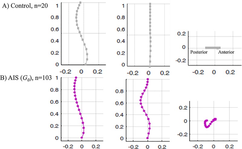
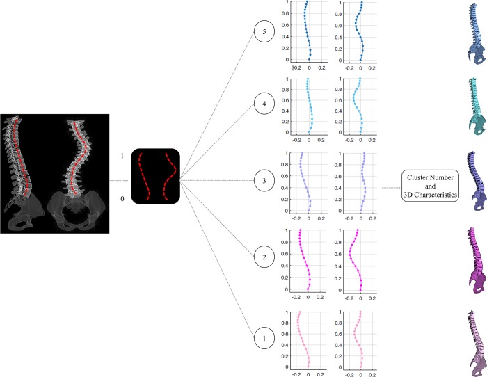
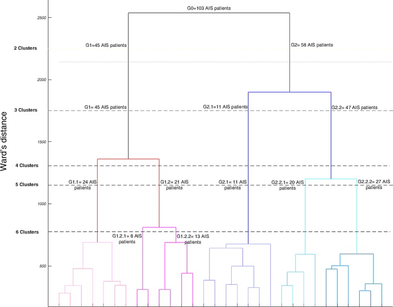
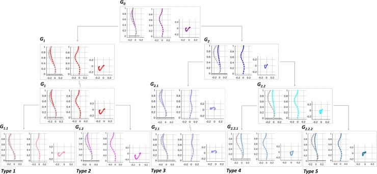
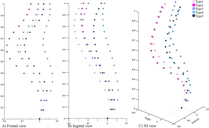
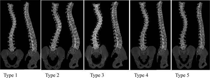
This study aimed to identify the differentiating parameters of the spinal curves' 2D projections through a hierarchical classification of the 3D spinal curve in adolescent idiopathic scoliosis (AIS). A total number of 103 right thoracic left lumbar pre-operative AIS patients were included retrospectively and consecutively. A total number of 20 non-scoliotic adolescents were included as the control group. All patients had biplanar X-rays and 3D reconstructions of the spine. The 3D spinal curve was calculated by interpolating the center of vertebrae and was isotropically normalized. A hierarchical classification of the normalized spinal curves was developed to group the patients based on the similarity of their 3D spinal curve. The spinal curves' 2D projections and clinical spinal measurements in the three anatomical planes were then statistically compared between these groups and between the scoliotic subtypes and the non-scoliotic controls. A total of 5 patient groups of right thoracic left lumbar AIS patients were identified. The characteristics of the posterior-anterior and sagittal views of the spines were: Type 1: Normal sagittal profile and S shape axial view. T1 is leveled or tilted to the right in the posterior view. Type 2: Hypokyphotic and a V shape axial view. T1 is tilted to the left in the posterior view. Type 3: Hypokyphotic (only T5-T10) and frontal imbalance, S shape axial view. T1 is leveled or tilted to the right, and 3 frontal curves. Type 4: Flat sagittal profile (T1-L2), slight frontal imbalance with a V shape axial view, T1 tilted to the left. Type 5: flat sagittal profile and forward trunk shift with a proximal kyphosis and S shape axial view. T1 is leveled or tilted to the right. In conclusion, a hierarchical classification of the 3D scoliotic spine allowed identifying various distinguishing features of the spinal curves in patients with a right thoracic curve in an orderly fashion. The subtypes' characteristics resulting from this 3D classification can be identified from the pairs of the frontal and sagittal spinal curves i.e. X-rays in right thoracic AIS patients.
本研究旨在通过对青少年特发性脊柱侧凸(AIS)的三维脊柱曲线进行分层分类,确定脊柱曲线二维投影的区分参数。回顾性连续纳入 103 例右侧胸弯左侧腰弯术前 AIS 患者。纳入 20 例非脊柱侧弯青少年作为对照组。所有患者均行双平面 X 线和脊柱三维重建。通过插值椎体中心计算三维脊柱曲线,并各向同性归一化。对归一化脊柱曲线进行分层分类,根据患者三维脊柱曲线的相似性对患者进行分组。然后在这些组之间以及脊柱侧弯亚型与非脊柱侧弯对照组之间比较脊柱曲线的二维投影和三个解剖平面的临床脊柱测量值。共确定了 5 组右侧胸弯左侧腰弯 AIS 患者。脊柱前后位和矢状位的特征如下:类型 1:矢状位正常,轴向观 S 形。后路 T1 右侧水平或倾斜。类型 2:胸椎后凸不足,轴向观 V 形。后路 T1 左侧倾斜。类型 3:胸椎后凸不足(仅 T5-T10)和冠状面不平衡,轴向观 S 形。T1 右侧水平或倾斜,有 3 个冠状面曲线。类型 4:矢状位平坦(T1-L2),轻度冠状面不平衡,轴向观 V 形,T1 左侧倾斜。类型 5:矢状位平坦,前躯干移位伴近端后凸和轴向观 S 形。T1 右侧水平或倾斜。总之,对三维脊柱侧弯进行分层分类可以有序地识别出右侧胸弯患者脊柱曲线的各种特征。从右胸弯 AIS 患者的 X 线可以识别出这种三维分类所产生的亚型特征,即前、矢状面脊柱曲线的组合。