Transplantation and Autoimmunity Laboratory, Rheumatology Department, University Hospital Marqués de Valdecilla-IDIVAL, Santander, Spain.
Faculty of Medicine, Rheumatology Department, University Hospital Marqués de Valdecilla-IDIVAL, Cantabria University, Santander, Spain.
Front Immunol. 2019 Mar 6;10:391. doi: 10.3389/fimmu.2019.00391. eCollection 2019.
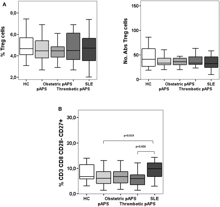
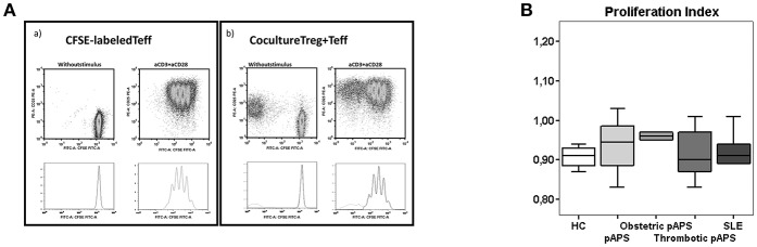
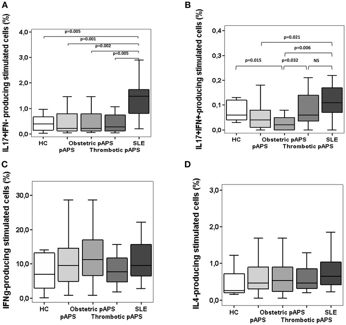
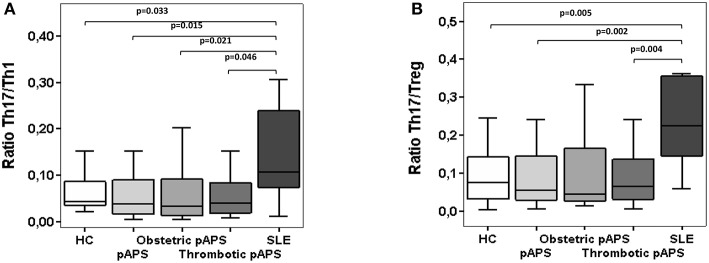
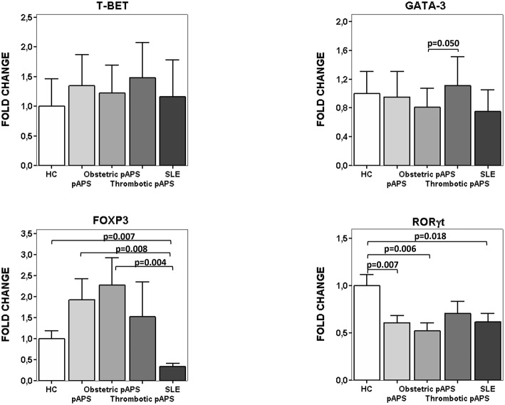
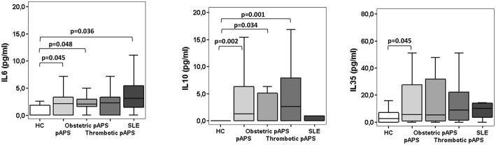
The role of the immune response in the pathogenesis of antiphospholipid syndrome (APS) remains elusive. It is possible that differences in the frequencies of Th17 cells and/or defects in the immunoregulatory mechanisms are involved in the pathogenesis of APS. Our aim was to determine the peripheral blood Th cells phenotype and the circulating cytokine profile in patients with primary APS (pAPS) and compare it with systemic lupus erythemathosus (SLE) as disease control group. The frequencies of circulating regulatory T cells (Tregs) were determined in PBMCs from 36 patients with pAPS by flow cytometry. As control groups we included 21 age- and gender-matched healthy controls (HC) and 11 patients with SLE. The suppressive capacity of Tregs was evaluated by coculture assay. On the other hand, intracellular cytokine production was assessed in Th1, Th2, and Th17 cells and circulating IL-6, IL-10, and IL-35 were measured by Cytometric Bead Array and ELISA. The quantification of Th master gene expression levels was performed by real time quantitative PCR. pAPS patients and SLE patients did not show differences in the percentage or number of Tregs compared to HC. The suppressive capacity of Tregs was also similar in the three study group. Instead, we found higher FoxP3·mRNA expression levels in pAPS patients and HC than SLE patients. Regarding the Th17 response, patients with pAPS and HC showed a significantly lower frequency of circulating Th17 cells than SLE. However, no differences were observed in the Th1 response between patients and controls. Thus, increased Th17/Th1 and Th17/Treg ratios were found in SLE patients but not in pAPS patients. pAPS and SLE patients had higher serum IL-6 levels than HC but there was not difference between both disease groups. Besides, a significant increase in the immunosuppressive cytokine levels was observed only in pAPS as compared to HC. Our data demonstrate an increased inflammatory profile of peripheral blood CD4 T cells from SLE as compared with pAPS mostly due to an increased Th17 response. In conclusion, there seems not to be a direct pathogenic role for Th cells in pAPS but in SLE.
免疫反应在抗磷脂综合征(APS)发病机制中的作用仍不清楚。可能是 Th17 细胞的频率差异和/或免疫调节机制的缺陷参与了 APS 的发病机制。我们的目的是确定原发性 APS(pAPS)患者的外周血 Th 细胞表型和循环细胞因子谱,并将其与系统性红斑狼疮(SLE)作为疾病对照组进行比较。通过流式细胞术确定 36 例 pAPS 患者 PBMC 中循环调节性 T 细胞(Tregs)的频率。作为对照组,我们纳入了 21 名年龄和性别匹配的健康对照(HC)和 11 名 SLE 患者。通过共培养试验评估 Tregs 的抑制能力。另一方面,通过流式细胞术评估 Th1、Th2 和 Th17 细胞内细胞因子的产生,并通过 Cytometric Bead Array 和 ELISA 测量循环 IL-6、IL-10 和 IL-35。通过实时定量 PCR 测定 Th 主基因表达水平的定量。与 HC 相比,pAPS 患者和 SLE 患者的 Tregs 百分比或数量没有差异。三个研究组的 Tregs 抑制能力也相似。相反,我们发现 pAPS 患者和 HC 的 FoxP3·mRNA 表达水平高于 SLE 患者。关于 Th17 反应,pAPS 患者和 HC 的循环 Th17 细胞频率明显低于 SLE 患者。然而,患者和对照组之间的 Th1 反应没有差异。因此,在 SLE 患者中发现了增加的 Th17/Th1 和 Th17/Treg 比值,但在 pAPS 患者中没有发现。pAPS 和 SLE 患者的血清 IL-6 水平高于 HC,但两组疾病之间无差异。此外,与 HC 相比,仅在 pAPS 中观察到免疫抑制细胞因子水平的显著增加。我们的数据表明,与 pAPS 相比,SLE 患者外周血 CD4 T 细胞的炎症谱增加,主要是由于 Th17 反应增加。总之,在 pAPS 中似乎没有 Th 细胞的直接致病作用,但在 SLE 中则有。