• 文献检索
  • 文档翻译
  • 深度研究
  • 学术资讯
  • Suppr Zotero 插件Zotero 插件
  • 邀请有礼
  • 套餐&价格
  • 历史记录
应用&插件
Suppr Zotero 插件Zotero 插件浏览器插件Mac 客户端Windows 客户端微信小程序
定价
高级版会员购买积分包购买API积分包
服务
文献检索文档翻译深度研究API 文档MCP 服务
关于我们
关于 Suppr公司介绍联系我们用户协议隐私条款
关注我们

Suppr 超能文献

核心技术专利:CN118964589B侵权必究
粤ICP备2023148730 号-1Suppr @ 2026

文献检索

告别复杂PubMed语法,用中文像聊天一样搜索,搜遍4000万医学文献。AI智能推荐,让科研检索更轻松。

立即免费搜索

文件翻译

保留排版,准确专业,支持PDF/Word/PPT等文件格式,支持 12+语言互译。

免费翻译文档

深度研究

AI帮你快速写综述,25分钟生成高质量综述,智能提取关键信息,辅助科研写作。

立即免费体验

退变性颈椎滑移患者的脊柱矢状位平衡和梯形畸形。

Spinal sagittal alignment and trapezoidal deformity in patients with degenerative cervical spondylolisthesis.

机构信息

Department of Orthopedic Surgery, Tokyo Medical University, 6-7-1 Nishishinjuku, Shinjuku-ku, Tokyo, Japan.

出版信息

Sci Rep. 2019 Mar 21;9(1):4992. doi: 10.1038/s41598-019-41079-3.

DOI:10.1038/s41598-019-41079-3
PMID:30899028
原文链接:https://pmc.ncbi.nlm.nih.gov/articles/PMC6428847/
Abstract

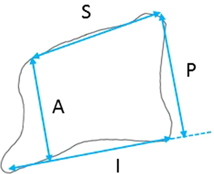
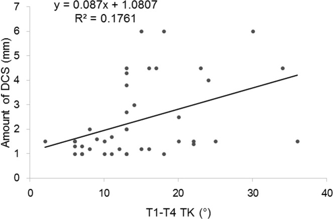
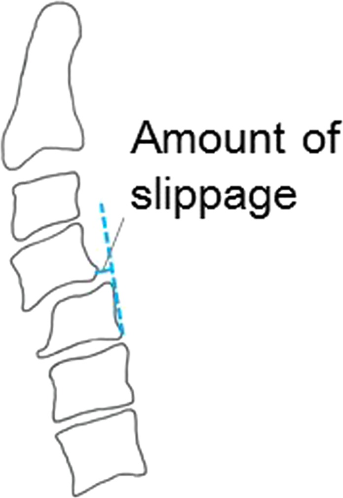
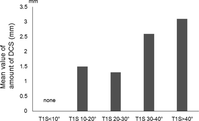
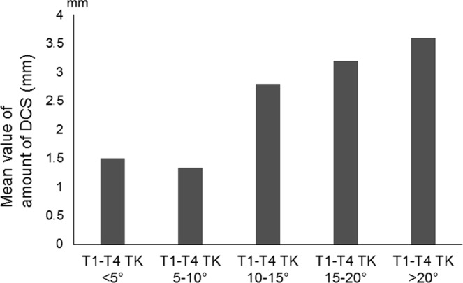
Degenerative cervical spondylolisthesis (DCS) is a cervical deformity arising from regressive changes where trapezoidal deformity characterized by hypertrophic osteophytes of spinal elements is often observed. There is a paucity of literature about the spinal contour of DCS and trapezoidal vertebrae. We conducted this research to clarify the relationship between spinal sagittal alignment and trapezoidal deformity in DCS. Total seventy-nine patients with cervical spondylosis were enrolled. Twenty-four patients who exhibited cervical spondylolisthesis were classified into DCS group. Other patients were classified into a control group. Measurements of radiographic parameters and trapezoidal deformity were made. DCS was found mostly in C3-C4 and C4-C5 (16 and 10 cases, respectively). T1S and T1-T4 TK was larger in the DCS group than in the control (T1S: 29.9 ± 2.3° vs. 23.7 ± 1.5°, T1-T4 TK: 14.9 ± 2.1° vs. 9.0 ± 1.4°). C2-C7A was smaller in DCS (3.5 ± 3.6° vs. 11.9 ± 2.3°). Trapezoidal deformity was apparent in the vertebra below the slipped segment. Among sagittal parameters, T1S and T1-T4 TK were positively correlated with DCS (r = 0.523 and r = 0.438, respectively). For these correlations with DCS, both logistic and linear regression models predicted threshold values of approximately 30° for T1S and 15° for T1-T4 TK responsible for DCS. DCS was mostly found in the middle cervical region. Among sagittal parameters, enlarged T1S and T1-T4 TK, which were strongly correlated with amount of slippage, was considered affected to DCS. Cervical kyphosis and trapezoidal deformity also exhibited strong correlations with DCS, and were considered responsible for clinical instability.

摘要

退行性颈椎失稳症(DCS)是一种颈椎畸形,源于退行性改变,常表现为颈椎各节段的梯形畸形,伴有骨赘形成。目前,关于 DCS 患者的脊柱矢状面形态与梯形椎体之间的关系,文献报道较少。本研究旨在明确 DCS 患者的脊柱矢状面形态与梯形椎体之间的关系。共纳入 79 例颈椎疾病患者。24 例颈椎滑脱患者被分为 DCS 组,其余患者被分为对照组。对两组患者的影像学参数和梯形变形情况进行测量。DCS 主要发生在 C3-C4 和 C4-C5(分别为 16 例和 10 例)。DCS 组患者的 T1S 和 T1-T4 TK 明显大于对照组(T1S:29.9±2.3° vs. 23.7±1.5°,T1-T4 TK:14.9±2.1° vs. 9.0±1.4°),C2-C7A 明显小于对照组(3.5±3.6° vs. 11.9±2.3°)。在滑脱节段以下的椎体中,可见明显的梯形变形。在矢状位参数中,T1S 和 T1-T4 TK 与 DCS 呈正相关(r=0.523 和 r=0.438)。对于与 DCS 相关的这些参数,logistic 和线性回归模型均预测 T1S 的阈值约为 30°,T1-T4 TK 的阈值约为 15°,与 DCS 相关。DCS 主要发生在颈椎中段。在矢状位参数中,与滑脱程度密切相关的 T1S 和 T1-T4 TK 增大,被认为与 DCS 有关。颈椎后凸和梯形变形也与 DCS 有很强的相关性,被认为与临床不稳定性有关。

https://cdn.ncbi.nlm.nih.gov/pmc/blobs/2ea0/6428847/b55c13fa5773/41598_2019_41079_Fig6_HTML.jpg
https://cdn.ncbi.nlm.nih.gov/pmc/blobs/2ea0/6428847/48692bd2091c/41598_2019_41079_Fig1_HTML.jpg
https://cdn.ncbi.nlm.nih.gov/pmc/blobs/2ea0/6428847/4e666429b2c8/41598_2019_41079_Fig2_HTML.jpg
https://cdn.ncbi.nlm.nih.gov/pmc/blobs/2ea0/6428847/7017566bd02d/41598_2019_41079_Fig3_HTML.jpg
https://cdn.ncbi.nlm.nih.gov/pmc/blobs/2ea0/6428847/ecd16461763a/41598_2019_41079_Fig4_HTML.jpg
https://cdn.ncbi.nlm.nih.gov/pmc/blobs/2ea0/6428847/c385801d0a5c/41598_2019_41079_Fig5_HTML.jpg
https://cdn.ncbi.nlm.nih.gov/pmc/blobs/2ea0/6428847/b55c13fa5773/41598_2019_41079_Fig6_HTML.jpg

相似文献

1
Spinal sagittal alignment and trapezoidal deformity in patients with degenerative cervical spondylolisthesis.退变性颈椎滑移患者的脊柱矢状位平衡和梯形畸形。
Sci Rep. 2019 Mar 21;9(1):4992. doi: 10.1038/s41598-019-41079-3.
2
Spinal Sagittal Alignment in Patients With Dropped Head Syndrome.患有垂头综合征患者的脊柱矢状面排列。
Spine (Phila Pa 1976). 2018 Nov 1;43(21):E1267-E1273. doi: 10.1097/BRS.0000000000002685.
3
Cervical sagittal alignment in adult hyperkyphosis treated by posterior instrumentation and in situ bending.采用后路内固定及原位折弯治疗成人脊柱后凸畸形时的颈椎矢状面排列
Orthop Traumatol Surg Res. 2017 Feb;103(1):53-59. doi: 10.1016/j.otsr.2016.10.003. Epub 2016 Nov 23.
4
Magnitude of preoperative cervical lordotic compensation and C2-T3 angle are correlated to increased risk of postoperative sagittal spinal pelvic malalignment in adult thoracolumbar deformity patients at 2-year follow-up.在成年胸腰椎畸形患者两年随访中,术前颈椎前凸代偿程度和C2-T3角与术后矢状位脊柱骨盆排列不齐风险增加相关。
Spine J. 2015 Aug 1;15(8):1756-63. doi: 10.1016/j.spinee.2015.04.007. Epub 2015 Apr 8.
5
Effect of cervical flexion and extension on thoracic sagittal alignment.颈椎屈伸对胸椎矢状面排列的影响。
J Orthop Surg (Hong Kong). 2019 Sep-Dec;27(3):2309499019876999. doi: 10.1177/2309499019876999.
6
Kinematic evaluation of cervical sagittal balance and thoracic inlet alignment in degenerative cervical spondylolisthesis using kinematic magnetic resonance imaging.使用运动磁共振成像对退变性颈椎滑脱症患者颈椎矢状面平衡和胸廓入口对线进行运动学评估。
Spine J. 2017 Sep;17(9):1272-1284. doi: 10.1016/j.spinee.2017.04.026. Epub 2017 Apr 26.
7
Sagittal Profile Response of Cervical Spine After Posterior Correction in Thoracic and Lumbar Adolescent Idiopathic Scoliosis: Correlation with Thoracic Kyphosis?青少年胸腰椎特发性脊柱侧凸后路矫正术后颈椎矢状面形态的变化:与胸椎后凸的相关性?
World Neurosurg. 2018 Dec;120:e333-e341. doi: 10.1016/j.wneu.2018.08.071. Epub 2018 Aug 23.
8
Trapezoidal Vertebral Body and Spine-Pelvis Sagittal Alignment in Patients with Lumber Spondylolisthesis.腰椎滑脱症患者的梯形椎体与脊柱-骨盆矢状位对线关系
Med Sci Monit. 2020 Nov 28;26:e927747. doi: 10.12659/MSM.927747.
9
Dimensions of the cervical neural foramen in conditions of spinal deformity: an ex vivo biomechanical investigation using specimen-specific CT imaging.脊柱畸形情况下颈椎神经孔的尺寸:一项使用特定标本CT成像的体外生物力学研究。
Eur Spine J. 2016 Jul;25(7):2155-65. doi: 10.1007/s00586-016-4409-4. Epub 2016 Feb 1.
10
Global sagittal spinal alignment at cervical flexion in patients with dropped head syndrome.患有垂头综合征患者颈椎前屈时的矢状位脊柱整体排列。
J Orthop Surg (Hong Kong). 2020 Sep-Dec;28(3):2309499020948266. doi: 10.1177/2309499020948266.

引用本文的文献

1
Cervical Spondylosis Diagnosis Based on Convolutional Neural Network with X-ray Images.基于 X 射线图像的卷积神经网络颈椎病诊断。
Sensors (Basel). 2024 May 26;24(11):3428. doi: 10.3390/s24113428.
2
The Posterolaterally Oriented and Laterally Downward Sloping Facet Joint Is a Risk Factor for Degenerative Cervical Spondylolisthesis and Myelopathy.后外侧方向且外侧向下倾斜的小关节是退行性颈椎滑脱和脊髓病的危险因素。
Spine Surg Relat Res. 2021 Dec 27;6(4):358-365. doi: 10.22603/ssrr.2021-0181. eCollection 2022.
3
Cervical facet joint effusion: A sign of instability in cervical degenerative spondylolisthesis.

本文引用的文献

1
T1 slope and degenerative cervical spondylolisthesis.T1 斜率与退变性颈椎滑脱
Spine (Phila Pa 1976). 2015 Feb 15;40(4):E220-6. doi: 10.1097/BRS.0000000000000722.
2
Risk factors for missed dynamic canal stenosis in the cervical spine.颈椎动态椎管狭窄漏诊的危险因素。
Spine (Phila Pa 1976). 2014 May 1;39(10):812-9. doi: 10.1097/BRS.0000000000000289.
3
Association of myelopathy scores with cervical sagittal balance and normalized spinal cord volume: analysis of 56 preoperative cases from the AOSpine North America Myelopathy study.
颈椎小关节积液:颈椎退行性椎体滑脱不稳的一个征象。
J Craniovertebr Junction Spine. 2022 Jan-Mar;13(1):38-41. doi: 10.4103/jcvjs.jcvjs_155_21. Epub 2022 Mar 9.
颈椎病评分与颈椎矢状平衡及脊髓体积比值的相关性分析:来自 AOSpine 北美颈椎病研究的 56 例术前病例分析。
Spine (Phila Pa 1976). 2013 Oct 15;38(22 Suppl 1):S161-70. doi: 10.1097/BRS.0b013e3182a7eb9e.
4
Prevalence and motion characteristics of degenerative cervical spondylolisthesis in the symptomatic adult.症状性成人退行性颈椎滑脱的患病率和运动特征。
Spine (Phila Pa 1976). 2013 Aug 1;38(17):E1115-20. doi: 10.1097/BRS.0b013e31829b1487.
5
The significance of facet joint cross-sectional area on magnetic resonance imaging in relationship to cervical degenerative spondylolisthesis.磁共振成像中关节突关节横截面积与颈椎退行性滑脱的关系。
Spine J. 2013 Aug;13(8):856-61. doi: 10.1016/j.spinee.2013.01.021. Epub 2013 Mar 7.
6
The natural history of degenerative spondylolisthesis of the cervical spine with 2- to 7-year follow-up.颈椎退行性脊椎滑脱症的自然病史:2 至 7 年随访结果。
Spine (Phila Pa 1976). 2013 Feb 15;38(4):E205-10. doi: 10.1097/BRS.0b013e31827de4fd.
7
Degenerative cervical spondylolisthesis: a systematic review.退行性颈椎滑脱:系统评价。
Int Orthop. 2011 Jun;35(6):869-75. doi: 10.1007/s00264-010-1203-5. Epub 2011 Jan 25.
8
Does vertebral level of pedicle subtraction osteotomy correlate with degree of spinopelvic parameter correction?经椎弓根截骨术的椎体水平与脊柱骨盆参数矫正程度是否相关?
J Neurosurg Spine. 2011 Feb;14(2):184-91. doi: 10.3171/2010.9.SPINE10129. Epub 2010 Dec 24.
9
Total sagittal spinal alignment in patients with lumbar canal stenosis accompanied by intermittent claudication.伴有间歇性跛行的腰椎管狭窄症患者的全脊柱矢状位排列。
Spine (Phila Pa 1976). 2010 Apr 20;35(9):E344-6. doi: 10.1097/BRS.0b013e3181c91121.
10
Degenerative spondylolisthesis does not influence surgical results of laminoplasty in elderly cervical spondylotic myelopathy patients.退变性脊椎滑脱不影响老年颈椎病患者接受椎板成形术的手术结果。
Eur Spine J. 2010 May;19(5):720-5. doi: 10.1007/s00586-010-1338-5. Epub 2010 Feb 27.