Wang Qinglei, Cui Yinghua, Lin Nan, Pang Shuchao
Intracardiac Intensive Care Unit, Affiliated Hospital of Jining Medical University, Jining, Shandong 272000, P.R. China.
Department of Cardiology (I), Affiliated Hospital of Jining Medical University, Jining, Shandong 272000, P.R. China.
Exp Ther Med. 2019 Apr;17(4):2741-2745. doi: 10.3892/etm.2019.7249. Epub 2019 Feb 7.
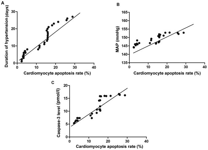
Correlation of cardiomyocyte apoptosis with duration of hypertension, severity of hypertension and caspase-3 expression in hypertensive rats was analyzed. Sixty male Sprague-Dawley (SD) rats were selected and randomly divided into the observation group (n=30) and control group (n=30), and the rat models of hypertension were established by virtue of transverse aortic constriction (TAC). The rats in the two groups were further divided into the 7-day subgroup (n=10), 14-day subgroup (n=10) and 28-day subgroup (n=10), respectively according to their survival time after TAC. The blood pressure values of the rats in each group were measured through intubation of carotid artery to calculate the mean arterial pressure (MAP). The conditions of cardiomyocyte apoptosis were detected using terminal dexynucleotidyl transferase-mediated dUTP nick end labeling (TUNEL) assay. Enzyme-linked immunosorbent assay (ELISA) was applied to measure the expression of caspase-3 in the myocardial tissues, and correlation analysis was performed. The MAPs in 7-, 14- and 28-day subgroups of the observation group were significantly higher than those in the corresponding subgroups of the control group (P<0.05). The 7-, 14- and 28-day subgroups of the observation group had remarkably elevated myocardial caspase-3 expression levels compared with the subgroups of the control group (P<0.05). The apoptosis rates of myocardial cells in the three subgroups of the observation group were obviously higher than those in the corresponding subgroups of the control group (P<0.05). Pearson's correlation analysis indicated that the cardiomyocyte apoptosis rate of hypertensive rats was positively correlated with the duration of hypertension, severity of hypertension and caspase-3 expression (P<0.05). Hypertension can induce apoptosis of myocardial cells, and the apoptosis becomes more serious with the constantly elevated level and prolonged duration of hypertension. In addition, the activity of caspase-3 has a close correlation with cardiomyocyte apoptosis.
分析高血压大鼠心肌细胞凋亡与高血压病程、高血压严重程度及半胱天冬酶-3表达的相关性。选取60只雄性Sprague-Dawley(SD)大鼠,随机分为观察组(n = 30)和对照组(n = 30),采用缩窄腹主动脉(TAC)法建立高血压大鼠模型。两组大鼠根据TAC术后存活时间进一步分为7天亚组(n = 10)、14天亚组(n = 10)和28天亚组(n = 10)。通过颈动脉插管测量各组大鼠血压值,计算平均动脉压(MAP)。采用末端脱氧核苷酸转移酶介导的dUTP缺口末端标记(TUNEL)法检测心肌细胞凋亡情况。应用酶联免疫吸附测定(ELISA)法检测心肌组织中半胱天冬酶-3的表达,并进行相关性分析。观察组7天、14天和28天亚组的MAP显著高于对照组相应亚组(P < 0.05)。观察组7天、14天和28天亚组心肌半胱天冬酶-3表达水平明显高于对照组亚组(P < 0.05)。观察组三个亚组心肌细胞凋亡率明显高于对照组相应亚组(P < 0.05)。Pearson相关性分析表明,高血压大鼠心肌细胞凋亡率与高血压病程、高血压严重程度及半胱天冬酶-3表达呈正相关(P < 0.05)。高血压可诱导心肌细胞凋亡,且随着高血压水平持续升高和病程延长,凋亡加重。此外,半胱天冬酶-3的活性与心肌细胞凋亡密切相关。