Research Institute of the McGill University Health Centre, Montreal, QC, Canada.
Centre for Host-Parasite Interactions, Institute of Parasitology, McGill University, Ste-Anne de Bellevue, QC, Canada.
Front Immunol. 2019 Mar 11;10:425. doi: 10.3389/fimmu.2019.00425. eCollection 2019.
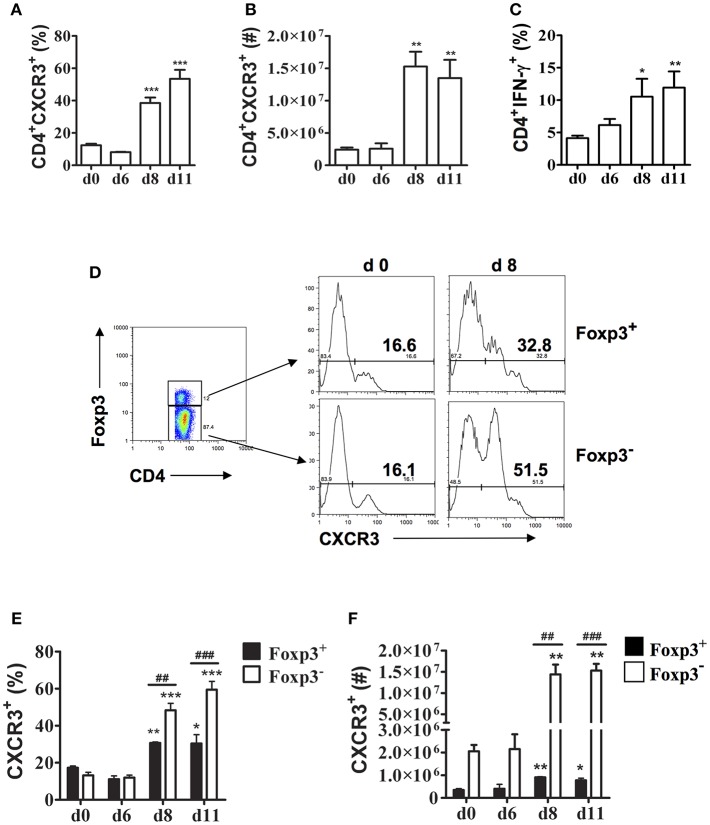
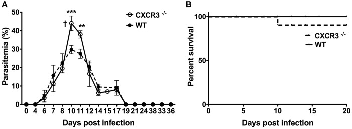
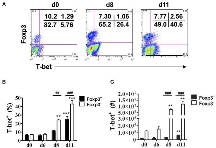
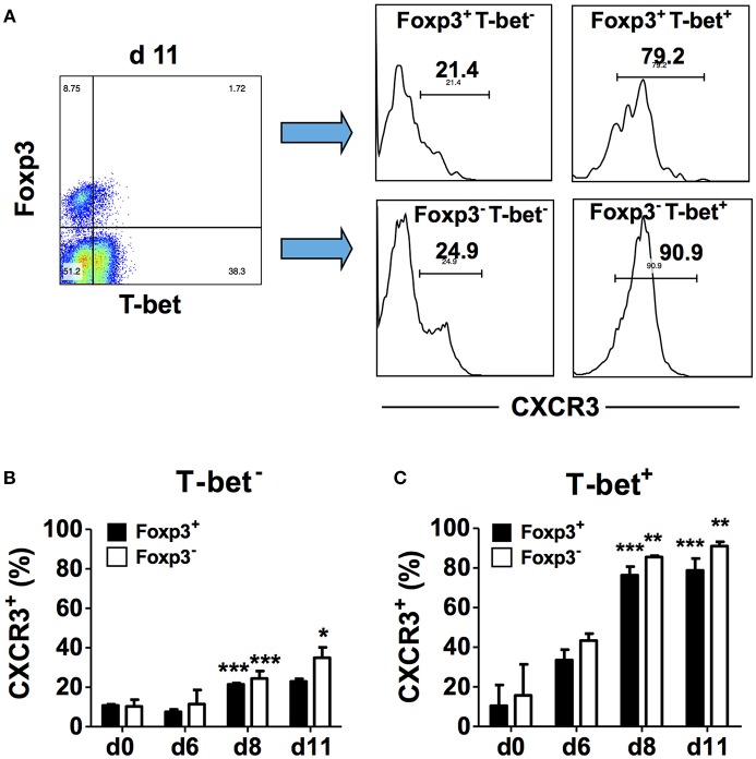
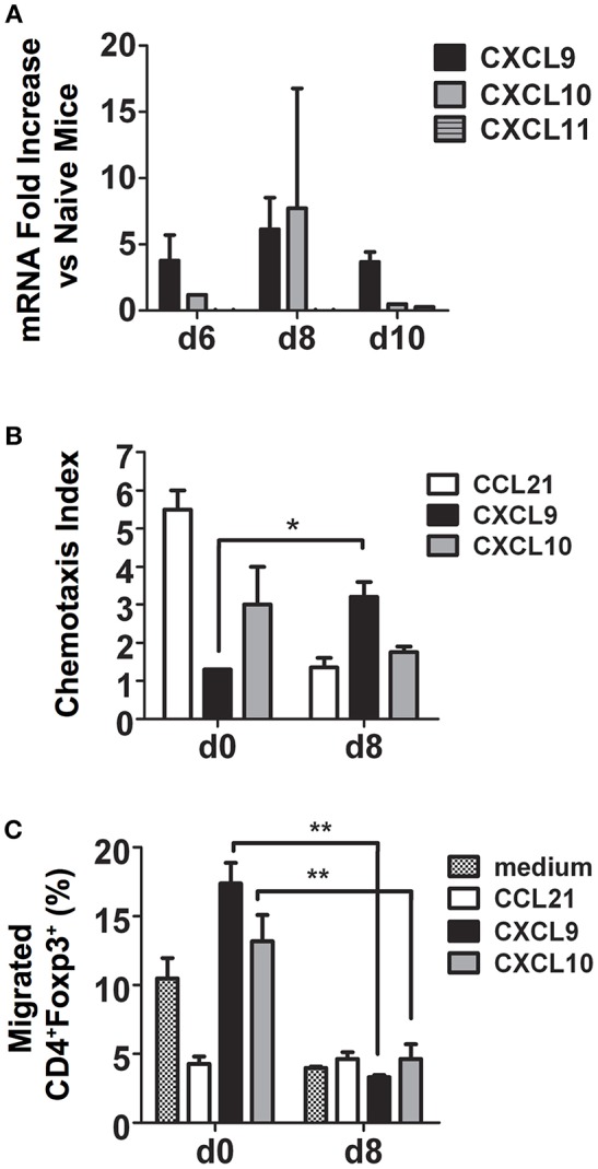
Control and elimination of blood-stage AS infection requires CD4 Th1 cells that secrete IFN-γ and T follicular help (Tfh) cells together with B cell production of antibody. Foxp3 regulatory T cells (Tregs) are also crucial to protect the host from immunopathology and severe disease, but these cells can suppress protective immune responses to malaria. The chemokine receptor CXCR3 expressed by activated T cells is important for trafficking of CD4 Th1 cells to sites of inflammation and infection. Previous studies demonstrated CXCR3 is expressed on CD4 T cells in the spleen during malaria, but the phenotype was not defined. We identified the phenotype of CD4 T cells that expressed CXCR3 in C57BL/6 (B6) mice during acute AS infection by analyzing expression of the transcription factors T-bet and Foxp3. We also investigated if CXCR3 contributes to control of parasite replication and survival. The frequency and number of CD4CXCR3 T cells increased dramatically in the spleen of infected B6 mice coincident with increased CD4IFN-γ T cells. CXCR3 was up-regulated on effector CD4Foxp3 T cells as well as Foxp3 Tregs. Consistent with our previous observations, CD4T-betFoxp3 T cells increased in B6 mice during acute infection. T-betFoxp3 Tregs also increased significantly and a high frequency of these cells expressed CXCR3 supporting the notion that these cells may be Th1-like Tregs. Despite this, the percentage of CD4Foxp3 Tregs from infected B6 mice that migrated to the CXCR3 ligands CXCL9 and CXCL10 was significantly less than naïve mice. To investigate the contribution of CXCR3 to control of acute blood-stage malaria, we compared the course and outcome of AS infection in wild-type (WT) B6 and CXCR3-deficient mice. Parasitemia levels were significantly higher around the time of peak parasitemia in CXCR3 compared to WT mice but survival was similar suggesting a role for CXCR3 in controlling parasite replication during acute AS infection. Together, our findings indicate Th1-like CD4T-betFoxp3 Tregs that express CXCR3 are induced during acute blood-stage malaria and suggest CXCR3 expression on CD4 Th1 cells may contribute to their migration to the spleen.
控制和消除血期疟原虫感染需要分泌 IFN-γ 的 CD4 Th1 细胞和滤泡辅助 T(Tfh)细胞,以及 B 细胞产生抗体。Foxp3 调节性 T 细胞(Tregs)对于保护宿主免受免疫病理和严重疾病也至关重要,但这些细胞可以抑制对疟疾的保护性免疫反应。激活的 T 细胞表达的趋化因子受体 CXCR3 对于 CD4 Th1 细胞向炎症和感染部位的迁移很重要。先前的研究表明,CXCR3 在疟疾期间表达于脾脏中的 CD4 T 细胞上,但表型尚未确定。我们通过分析转录因子 T-bet 和 Foxp3 的表达,鉴定了 C57BL/6(B6)小鼠在急性疟原虫感染期间表达 CXCR3 的 CD4 T 细胞的表型。我们还研究了 CXCR3 是否有助于控制寄生虫复制和存活。在感染 B6 小鼠的脾脏中,CD4CXCR3 T 细胞的频率和数量急剧增加,同时 CD4IFN-γ T 细胞也增加。CXCR3 在效应 CD4Foxp3 T 细胞和 Foxp3 Tregs 上上调。与我们之前的观察结果一致,在急性感染期间,B6 小鼠中的 CD4T-betFoxp3 T 细胞增加。T-betFoxp3 Tregs 也显著增加,这些细胞中的高频率表达 CXCR3,支持这些细胞可能是 Th1 样 Tregs 的观点。尽管如此,从感染 B6 小鼠中迁移到 CXCR3 配体 CXCL9 和 CXCL10 的 CD4Foxp3 Tregs 的百分比明显低于未感染的小鼠。为了研究 CXCR3 对控制急性血期疟疾的贡献,我们比较了野生型(WT)B6 和 CXCR3 缺陷型小鼠中疟原虫感染的过程和结果。与 WT 小鼠相比,CXCR3 中的寄生虫血症水平在寄生虫血症峰值周围显着升高,但存活率相似,表明 CXCR3 在控制急性疟原虫感染期间的寄生虫复制中发挥作用。总之,我们的发现表明,在急性血期疟疾期间诱导表达 CXCR3 的 Th1 样 CD4T-betFoxp3 Tregs,并表明 CD4 Th1 细胞上的 CXCR3 表达可能有助于其向脾脏的迁移。