Mochizuki Tomoharu, Koga Yoshio, Tanifuji Osamu, Sato Takashi, Watanabe Satoshi, Koga Hiroshi, Kobayashi Koichi, Omori Go, Endo Naoto
Division of Orthopaedic Surgery, Department of Regenerative and Transplant Medicine, Niigata University Graduate School of Medical and Dental Science, 1-757 Asahimachi-dori Chuo-ku, Niigata, 951-8510, Japan.
Department of Orthopaedic Surgery, Nioji Onsen Hospital, Niigata, Japan.
J Exp Orthop. 2019 Mar 28;6(1):14. doi: 10.1186/s40634-019-0180-x.
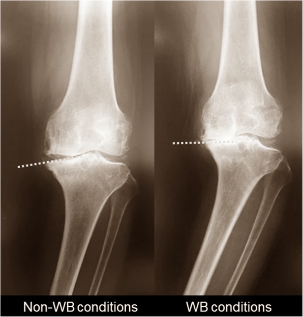
The inclination of the medial compartment of the proximal tibia (MCT) is assumed to be a critical factor for varus alignment in advanced knee osteoarthritis (OA). This study was aimed at investigating; (1) whether the inclination of MCT is aligned parallel to the ground under weight-bearing (WB) conditions; (2) whether this is associated with the change in alignment and the relative position between the bones; and (3) whether the tibia or femur mainly contributes to the changes.
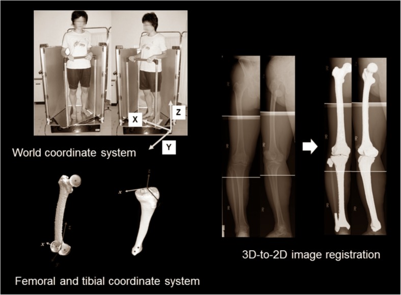
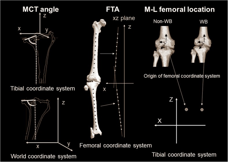
We examined 102 knees (84 women, 18 men; mean 75 years). A three-dimensional (3D) assessment system was applied on biplanar whole lower extremity radiographies using 3D-to-2D image registration technique. The evaluation parameters were 1) MCT angle, 2) femorotibial angle (FTA), 3) medial-lateral femoral location to the tibia (M-L femoral location), 4) WB line passing point, and 5) tibial position to WB line (tibial position) and 6) femoral postion to WB line (femoral position). Each parameter was evaluated in non-WB and WB conditions, and the differences (Δ-parameters).
MCT angle in the world coordinate system was larger than that in the tibial coordinate system (p < 0.0001). ΔMCT angle was correlated with ΔFTA (p = 0.002) and ΔM-L femoral location (p = 0.004). The tibial position was the more dominant factor for ΔMCT angle (p = 0.001), ΔFTA (p < 0.0001), and ΔWB line passing point (p < 0.0001) .
The inclination in MCT was aligned parallel to the ground under WB conditions (tibial parallel phenomenon). The parallel phenomenon was associated with the change of alignment and the relative position between the bones in the coronal plane. These phenomena were produced mainly by the tibia, not the femur.
Level IV.
胫骨近端内侧间室(MCT)的倾斜度被认为是晚期膝关节骨关节炎(OA)内翻对线的关键因素。本研究旨在调查:(1)在负重(WB)条件下,MCT的倾斜度是否与地面平行;(2)这是否与对线变化及骨骼间的相对位置有关;(3)胫骨或股骨对这些变化的主要贡献程度。
我们检查了102个膝关节(84名女性,18名男性;平均年龄75岁)。使用三维到二维图像配准技术,在双平面全下肢X线片上应用三维(3D)评估系统。评估参数包括:1)MCT角,2)股胫角(FTA),3)股骨相对于胫骨的内外侧位置(M-L股骨位置),4)负重线通过点,5)胫骨相对于负重线的位置(胫骨位置),以及6)股骨相对于负重线的位置(股骨位置)。在非负重和负重条件下对每个参数进行评估,并计算差异(Δ参数)。
世界坐标系中的MCT角大于胫骨坐标系中的MCT角(p < 0.0001)。ΔMCT角与ΔFTA(p = 0.002)和ΔM-L股骨位置(p = 0.004)相关。胫骨位置是ΔMCT角(p = 0.001)、ΔFTA(p < 0.0001)和Δ负重线通过点(p < 0.0001)的更主要影响因素。
在负重条件下,MCT的倾斜度与地面平行(胫骨平行现象)。这种平行现象与冠状面内的对线变化及骨骼间的相对位置有关。这些现象主要由胫骨而非股骨产生。
IV级。