Yin Guo Nan, Ock Jiyeon, Choi Min Ji, Song Kang Moon, Ghatak Kalyan, Minh Nguyen Nhat, Kwon Mi Hye, Seong Do Hwan, Jin Hai Rong, Ryu Ji Kan, Suh Jun Kyu
National Research Center for Sexual Medicine and Department of Urology, Inha University School of Medicine, Incheon, Korea.
Department of Urology, Yantai Yuhuangding Hospital Affiliated to Medical College of Qingdao University, Yantai, China.
World J Mens Health. 2020 Jan;38(1):123-131. doi: 10.5534/wjmh.180091. Epub 2019 Mar 15.
To establish a simple and nonenzymatic technique to isolate endothelial cells (ECs) and pericytes from human corpus cavernosum tissue and to evaluate the angiogenic ability of the human cavernous EC or pericytes for the study of high glucose-induced angiopathy.
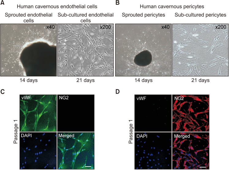
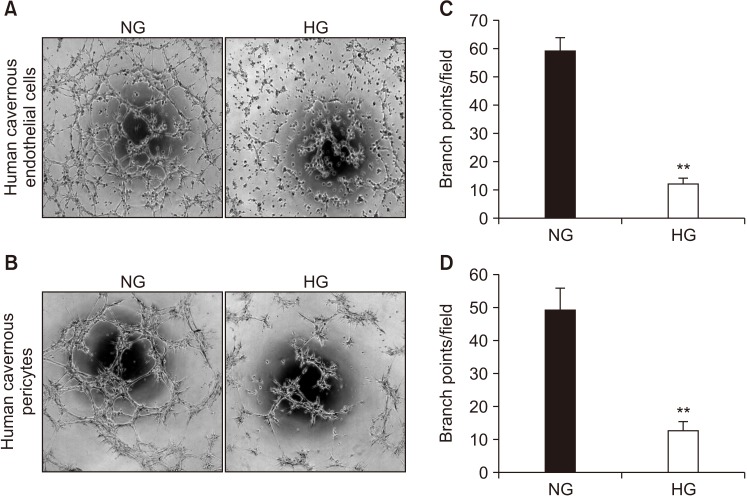
For primary human cavernous EC culture, cavernous tissues were implanted into Matrigel in dishes. For primary human cavernous pericyte culture, cavernous tissues were settled by gravity into dishes. We performed immunocytochemistry and Western blot to determine phenotype and morphologic changes from passage 1 to 5. The primary cultured cells were exposed to a normal-glucose (5 mmol/L) or a high-glucose (30 mmol/L) condition, and then tube formation assay was done.
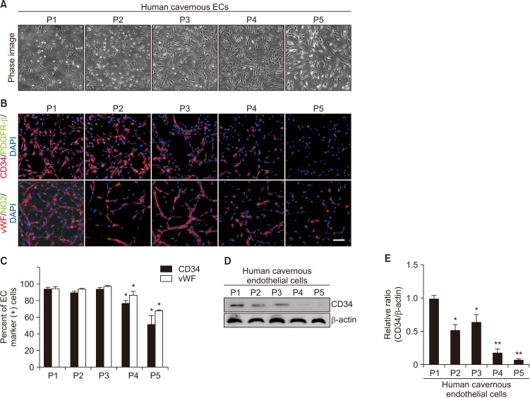
We successfully isolated high-purity EC and pericytes from human corpus cavernosum tissue. Primary cultured EC showed highly positive staining for von Willebrand factor, and pericyte revealed positive staining for NG2 and platelet-derived growth factor receptor-β. Primary cultured EC and pericytes maintained their cellular characteristics up to passage 2 or 3. However, we observed significant changes in their typical phenotype from the passage 4 and morphological characteristics from the passage 3. Human cavernous EC or pericytes formed well-organized capillary-like structures in normal-glucose condition, whereas severely impaired tube formation was detected in high-glucose condition.
This study provides a simple and nonenzymatic method for primary culture of human cavernous EC and pericytes. Our study will aid us to understand the pathophysiology of diabetic erectile dysfunction, and also be a valuable tool for determining the efficacy of candidate therapeutic targets.
建立一种简单的非酶技术,从人阴茎海绵体组织中分离内皮细胞(ECs)和平滑肌周细胞,并评估人海绵体EC或周细胞的血管生成能力,以研究高糖诱导的血管病变。
对于原代人海绵体EC培养,将海绵体组织植入培养皿中的基质胶内。对于原代人海绵体周细胞培养,将海绵体组织靠重力沉降到培养皿中。我们进行免疫细胞化学和蛋白质印迹分析,以确定第1代到第5代细胞的表型和形态变化。将原代培养的细胞置于正常葡萄糖(5 mmol/L)或高糖(30 mmol/L)条件下,然后进行管腔形成试验。
我们成功地从人阴茎海绵体组织中分离出高纯度的EC和周细胞。原代培养的EC对血管性血友病因子呈高度阳性染色,周细胞对NG2和血小板衍生生长因子受体-β呈阳性染色。原代培养的EC和周细胞在传代至第2代或第3代时仍保持其细胞特征。然而,我们观察到从第4代起其典型表型有显著变化,从第3代起其形态特征有显著变化。人海绵体EC或周细胞在正常葡萄糖条件下形成组织良好的毛细血管样结构,而在高糖条件下检测到管腔形成严重受损。
本研究提供了一种简单的非酶方法用于原代培养人海绵体EC和周细胞。我们的研究将有助于我们了解糖尿病性勃起功能障碍的病理生理学,也是确定候选治疗靶点疗效的有价值工具。