Department of Radiology, Severance Hospital, Research Institute of Radiological Science, Yonsei University College of Medicine, Seoul, South Korea.
Department of Pediatrics, Institute of Allergy, Brain Korea 21 PLUS Project for Medical Science, Severance Children's Hospital, Yonsei University College of Medicine, Seoul, South Korea.
PLoS One. 2019 Apr 1;14(4):e0214647. doi: 10.1371/journal.pone.0214647. eCollection 2019.
To investigate the feasibility of CT-based quantitative airway and air-trapping measurements and to assess their correlation with pulmonary function in children with post-infectious bronchiolitis obliterans (PIBO).
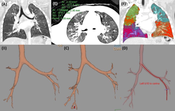
This retrospective study approved by the institutional review board included chest CT scans and pulmonary function tests (PFT) completed between January 2005 and December 2016 in children diagnosed with PIBO. The quantitative analysis of segmental and subsegmental bronchi was performed on each chest CT scan, measuring the areas or diameters of lumens, walls, or the entire airway. The air-trapping volume (ATV), the volume of lung area exhibiting lower attenuation than the mean attenuation of normal and air-trapping areas, was also measured in each lobe. Comparison analyses between CT parameters and PFT results were performed with Pearson or Spearman correlation.
In total, 23 patients were enrolled (mean age 7.0 ± 3.3 years; range, 4-15 years). We successfully measured 89.6% of all segmental bronchi. In the airway analysis, wall area showed a negative correlation with forced expiratory volume in one second (FEV1) in the majority of the pulmonary lobes. Air-trapping analyses demonstrated that ATV was negatively correlated with FEV1 and positively correlated with reactance at 5 Hz.
Quantitative airway and air-trapping measurements from chest CT are feasible and correlate with pulmonary function in pediatric PIBO patients.
研究基于 CT 的定量气道和空气潴留测量的可行性,并评估其与感染后细支气管炎闭塞(PIBO)患儿的肺功能的相关性。
本回顾性研究经机构审查委员会批准,纳入了 2005 年 1 月至 2016 年 12 月间在诊断为 PIBO 的患儿中完成的胸部 CT 扫描和肺功能检查(PFT)。对每例胸部 CT 扫描进行节段性和亚节段性支气管的定量分析,测量管腔、管壁或整个气道的面积或直径。在每个肺叶中还测量了空气潴留量(ATV),即比正常和空气潴留区域平均衰减值低的肺区衰减体积。采用 Pearson 或 Spearman 相关分析进行 CT 参数与 PFT 结果之间的比较分析。
共纳入 23 例患者(平均年龄 7.0 ± 3.3 岁;范围,4-15 岁)。我们成功测量了所有节段性支气管的 89.6%。在气道分析中,大多数肺叶的管壁面积与第一秒用力呼气量(FEV1)呈负相关。空气潴留分析表明,ATV 与 FEV1 呈负相关,与 5Hz 电抗呈正相关。
胸部 CT 的定量气道和空气潴留测量是可行的,与小儿 PIBO 患者的肺功能相关。