From the School of Cardiovascular Medicine and Sciences, King's College London British Heart Foundation Centre, United Kingdom (W.G., Z.N., J.D., Z.Z., Y.H., Q.X.).
Key Laboratory of Pulmonary Vascular Medicine and FuWai Hospital, State Key Laboratory of Cardiovascular Disease, Peking Union Medical College, Chinese Academy of Medical Sciences, Beijing (Y.-Q.T., S.-J.Z., Z.-C.L., X.-J.W., Z.-C.J.).
Arterioscler Thromb Vasc Biol. 2019 Jun;39(6):1055-1071. doi: 10.1161/ATVBAHA.119.312399.
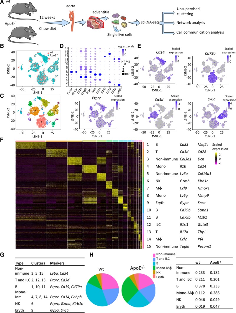
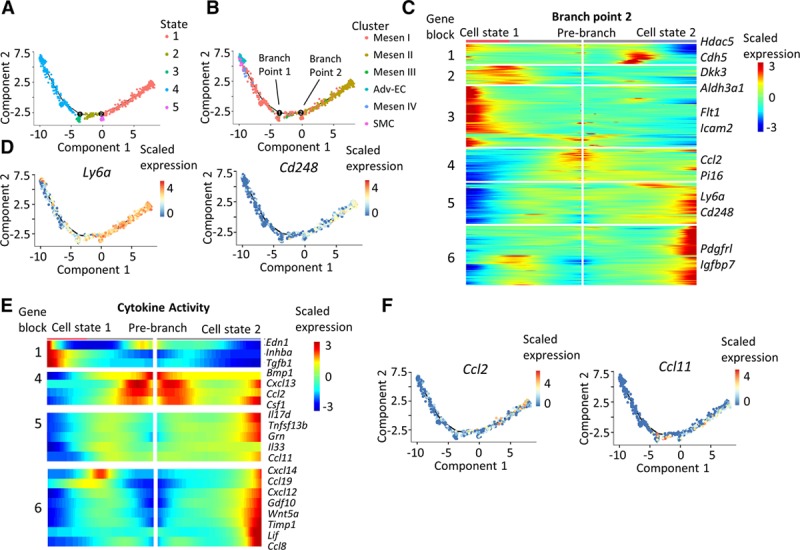
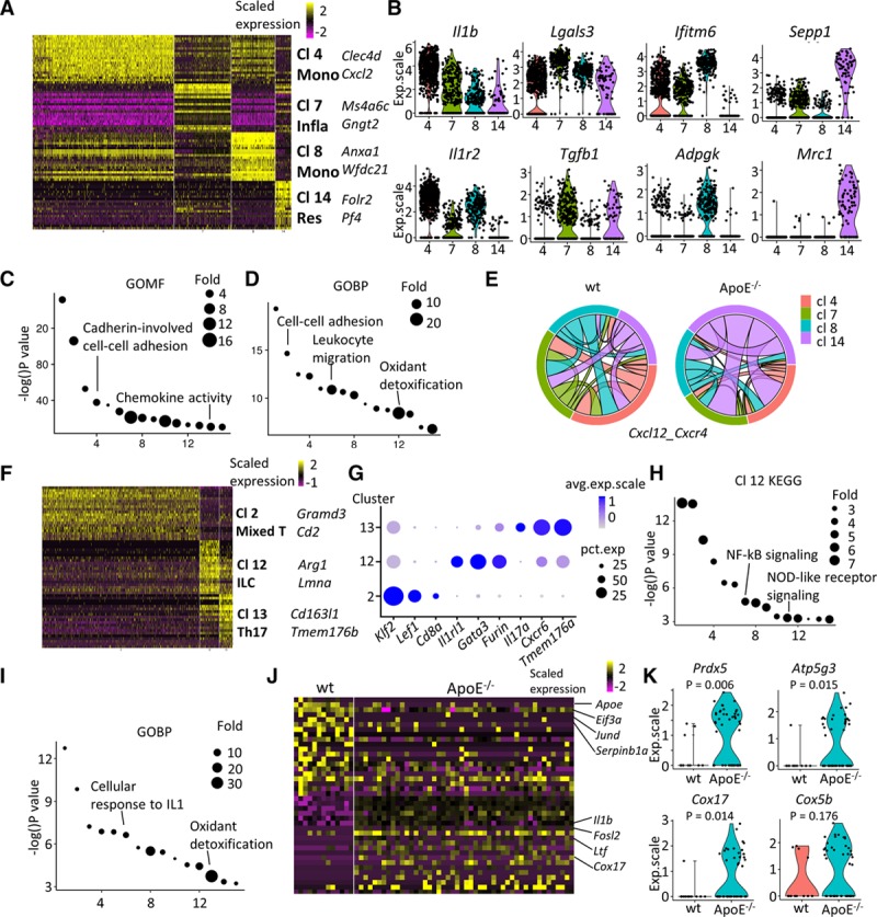
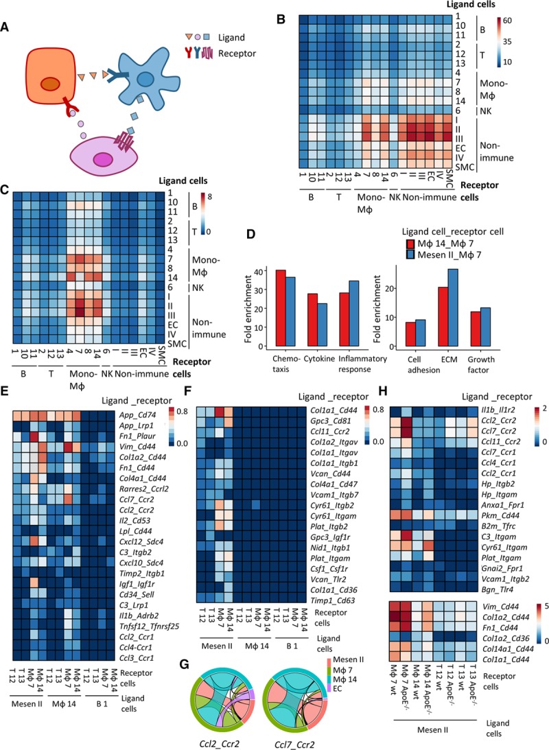
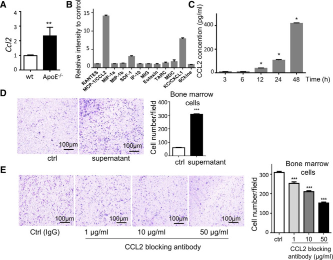
Objective- Vascular adventitia encompasses progenitors and is getting recognized as the major site of inflammation in early stage of atherosclerosis. However, the cellular atlas of the heterogeneous adventitial cells, the intercellular communication, the cellular response of adventitia to hyperlipidemia, and its contribution to atherosclerosis have been elusive. Approach and Results- Single-cell RNA sequencing was applied to wt (wild type) and ApoE (apolipoprotein E)-deficient aortic adventitia from 12-week-old C57BL/6J mice fed on normal laboratory diet with early stage of atherosclerosis. Unbiased clustering analysis revealed that the landscape of adventitial cells encompassed adventitial mesenchyme cells, immune cells (macrophages, T cells, and B cells), and some types of rare cells, for example, neuron, lymphatic endothelial cells, and innate lymphoid cells. Seurat clustering analysis singled out 6 nonimmune clusters with distinct transcriptomic profiles, in which there predominantly were stem/progenitor cell-like and proinflammatory population (Mesen II). In ApoE-deficient adventitia, resident macrophages were activated and related to increased myeloid cell infiltration in the adventitia. Cell communication analysis further elucidated enhanced interaction between a mesenchyme cluster and inflammatory macrophages in ApoE-deficient adventitia. In vitro transwell assay confirmed the proinflammatory role of SCA1 (stem cell antigen 1 positive) Mesen II population with increased CCL2 (chemokine [C-C motif] ligand 2) secretion and thus increased capacity to attract immune cells in ApoE-deficient adventitia. Conclusions- Cell atlas defined by single-cell RNA sequencing depicted the heterogeneous cellular landscape of the adventitia and uncovered several types of cell populations. Furthermore, resident cell interaction with immune cells appears crucial at the early stage of atherosclerosis.
目的-血管外膜包含祖细胞,并被认为是动脉粥样硬化早期炎症的主要部位。然而,异质性外膜细胞的细胞图谱、细胞间通讯、外膜细胞对高脂血症的反应及其对动脉粥样硬化的贡献仍不清楚。
方法和结果-应用单细胞 RNA 测序技术对 12 周龄 C57BL/6J 小鼠的野生型(wt)和载脂蛋白 E 缺陷(ApoE)主动脉外膜进行分析,这些小鼠在正常实验室饮食下喂养,处于动脉粥样硬化的早期阶段。无偏聚类分析显示,外膜细胞的景观包括外膜间充质细胞、免疫细胞(巨噬细胞、T 细胞和 B 细胞)和一些类型的稀有细胞,例如神经元、淋巴内皮细胞和固有淋巴细胞。Seurat 聚类分析单独鉴定出 6 个具有不同转录组特征的非免疫簇,其中主要是干细胞/祖细胞样和促炎群体(Mesen II)。在 ApoE 缺陷的外膜中,驻留巨噬细胞被激活,并与外膜中髓样细胞浸润的增加有关。细胞通讯分析进一步阐明了 ApoE 缺陷的外膜中间充质簇与炎症巨噬细胞之间增强的相互作用。体外 Transwell 实验证实了 SCA1(干细胞抗原 1 阳性)Mesen II 群体的促炎作用,其 CCL2(趋化因子[C-C 基序]配体 2)分泌增加,从而在外膜缺陷的 ApoE 中吸引免疫细胞的能力增强。
结论-单细胞 RNA 测序定义的细胞图谱描绘了外膜的异质细胞景观,并揭示了几种细胞群体。此外,驻留细胞与免疫细胞的相互作用在动脉粥样硬化的早期阶段似乎至关重要。