Nash Family Department of Neuroscience, Friedman Brain Institute, Icahn School of Medicine at Mount Sinai, New York, NY, USA.
Department of Genetics and Genomic Sciences, Icahn Institute of Genomics and Multiscale Biology, Icahn School of Medicine at Mount Sinai, New York, NY, USA.
EBioMedicine. 2019 Apr;42:252-269. doi: 10.1016/j.ebiom.2019.03.064. Epub 2019 Apr 3.
Glioblastoma (GBM), a highly malignant brain tumor, invariably recurs after therapy. Quiescent GBM cells represent a potential source of tumor recurrence, but little is known about their molecular underpinnings.
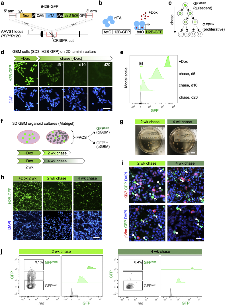
Patient-derived GBM cells were engineered by CRISPR/Cas9-assisted knock-in of an inducible histone2B-GFP (iH2B-GFP) reporter to track cell division history. We utilized an in vitro 3D GBM organoid approach to isolate live quiescent GBM (qGBM) cells and their proliferative counterparts (pGBM) to compare stem cell properties and therapy resistance. Gene expression programs of qGBM and pGBM cells were analyzed by RNA-Seq and NanoString platforms.
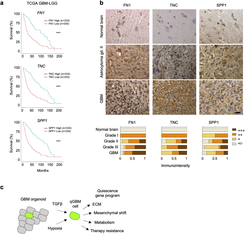
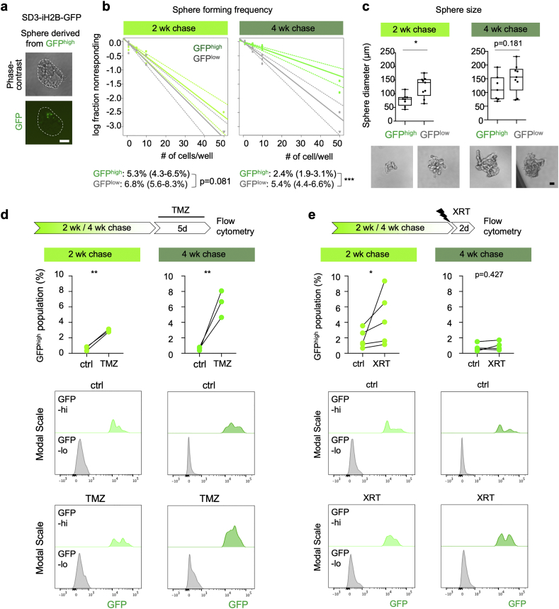
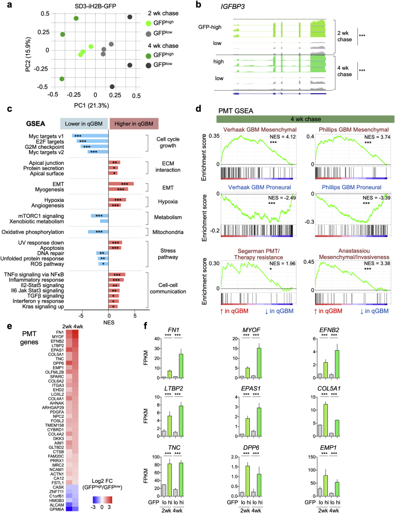
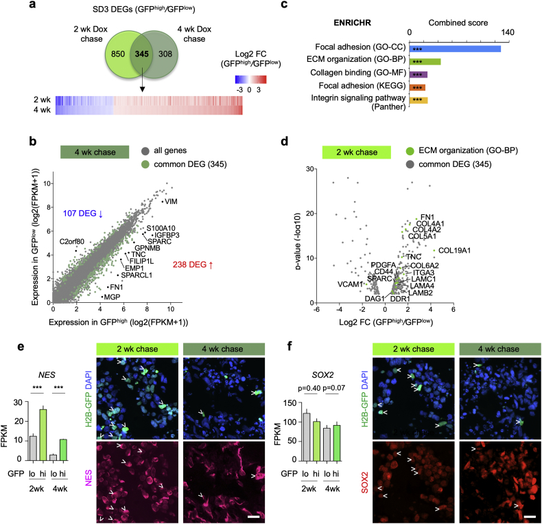
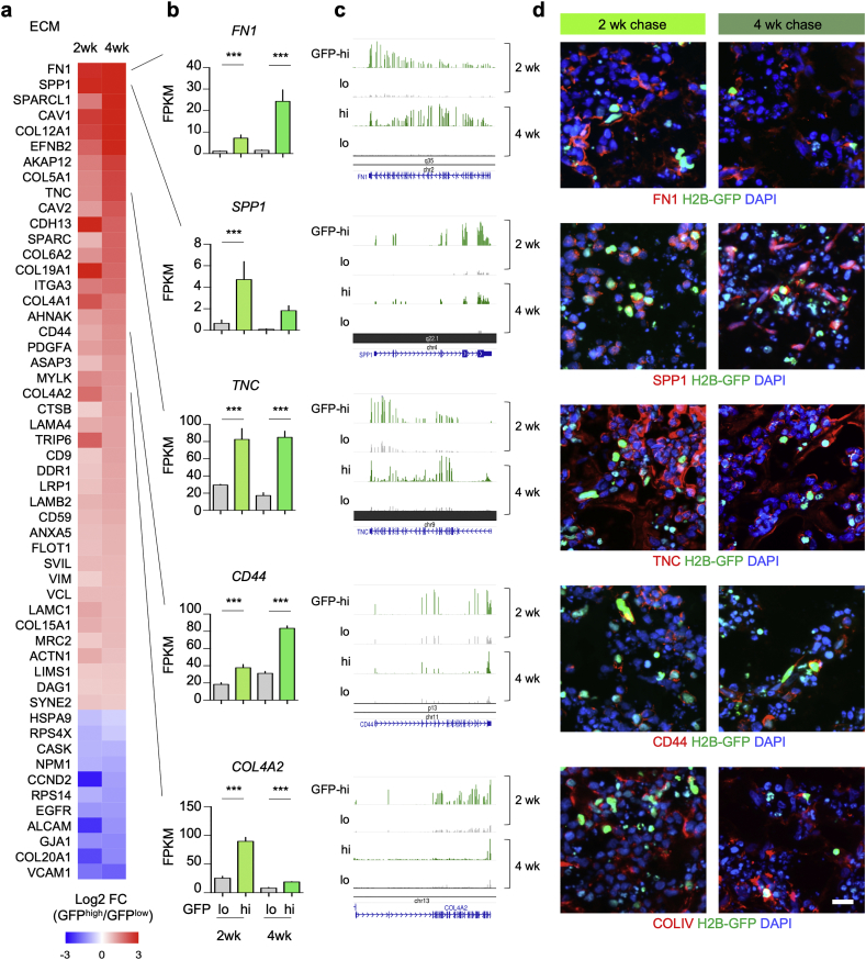
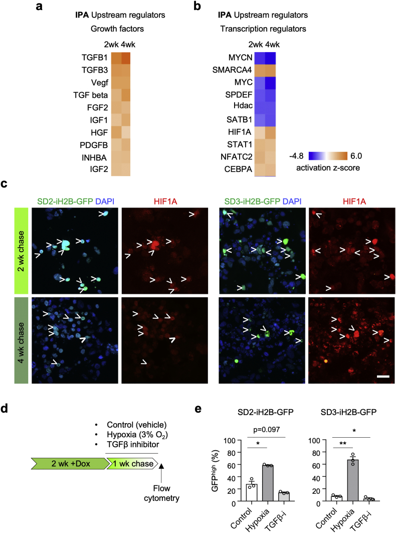
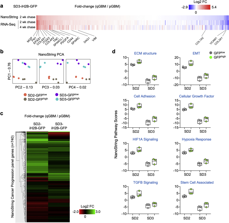
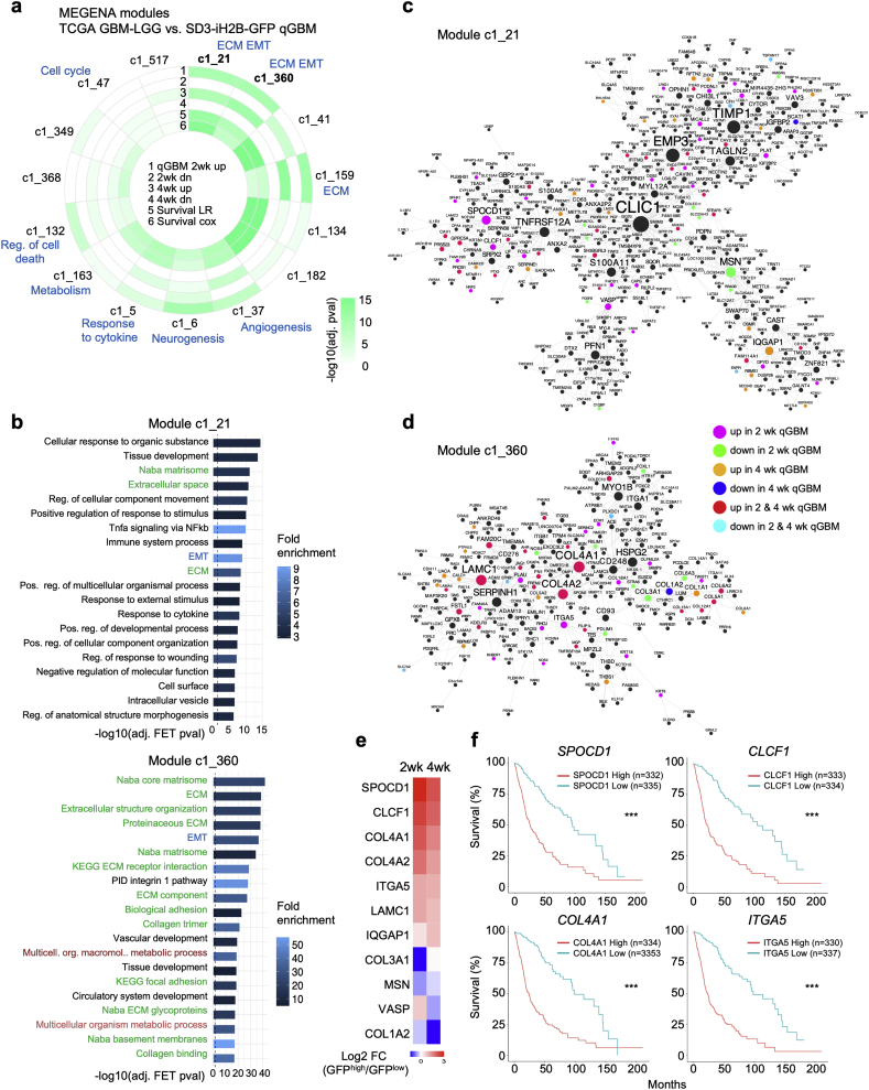
H2B-GFP-retaining qGBM cells exhibited comparable self-renewal capacity but higher therapy resistance relative to pGBM. Quiescent GBM cells expressed distinct gene programs that affect cell cycle control, metabolic adaptation, and extracellular matrix (ECM) interactions. Transcriptome analysis also revealed a mesenchymal shift in qGBM cells of both proneural and mesenchymal GBM subtypes. Bioinformatic analyses and functional assays in GBM organoids established hypoxia and TGFβ signaling as potential niche factors that promote quiescence in GBM. Finally, network co-expression analysis of TCGA glioma patient data identified gene modules that are enriched for qGBM signatures and also associated with survival rate.
Our in vitro study in 3D GBM organoids supports the presence of a quiescent cell population that displays self-renewal capacity, high therapy resistance, and mesenchymal gene signatures. It also sheds light on how GBM cells may acquire and maintain quiescence through ECM organization and interaction with niche factors such as TGFβ and hypoxia. Our findings provide a starting point for developing strategies to tackle the quiescent population of GBM. FUND: National Institutes of Health (NIH) and Deutsche Forschungsgemeinschaft (DFG).
胶质母细胞瘤(GBM)是一种高度恶性的脑肿瘤,在治疗后总会复发。静止的 GBM 细胞代表了肿瘤复发的潜在来源,但人们对其分子基础知之甚少。
通过 CRISPR/Cas9 辅助的诱导型组蛋白 2B-GFP(iH2B-GFP)报告基因敲入,对患者来源的 GBM 细胞进行工程改造,以跟踪细胞分裂史。我们利用体外 3D GBM 类器官方法分离活的静止 GBM(qGBM)细胞及其增殖对应物(pGBM),以比较干细胞特性和治疗耐药性。通过 RNA-Seq 和 NanoString 平台分析 qGBM 和 pGBM 细胞的基因表达程序。
H2B-GFP 保留的 qGBM 细胞表现出相当的自我更新能力,但相对于 pGBM 细胞具有更高的治疗耐药性。静止的 GBM 细胞表达了影响细胞周期控制、代谢适应和细胞外基质(ECM)相互作用的独特基因程序。转录组分析还揭示了神经前体细胞和间充质 GBM 亚型的 qGBM 细胞发生了间质转化。GBM 类器官中的生物信息学分析和功能测定确定了缺氧和 TGFβ 信号作为促进 GBM 静止的潜在龛位因素。最后,TCGA 神经胶质瘤患者数据的网络共表达分析确定了富含 qGBM 特征的基因模块,并且与存活率相关。
我们在 3D GBM 类器官中的体外研究支持存在具有自我更新能力、高治疗耐药性和间质基因特征的静止细胞群体。它还揭示了 GBM 细胞如何通过 ECM 组织和与 TGFβ 和缺氧等龛位因素的相互作用来获得和维持静止状态。我们的发现为开发策略以解决 GBM 的静止群体提供了一个起点。
美国国立卫生研究院(NIH)和德国研究基金会(DFG)。