Department of Vascular Surgery, Changhai Hospital, Second Military Medical University, Shanghai, China.
College of Animal Science and Technology, Anhui Agricultural University, Hefei, China.
Contrast Media Mol Imaging. 2019 Mar 6;2019:5940301. doi: 10.1155/2019/5940301. eCollection 2019.
The aim of this study was to evaluate the potential of microcomputed tomography (micro-CT) using the intravascular contrast agent ExiTron nano 12000 for aorta imaging and monitoring the dynamic changing process of the aorta in mouse models with aortic aneurysm and dissection.
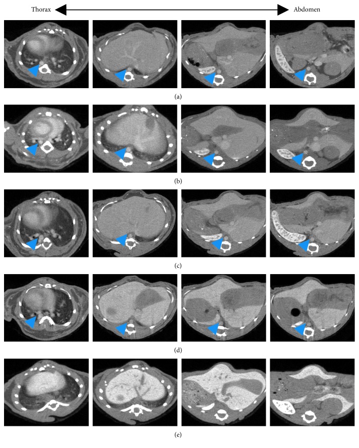
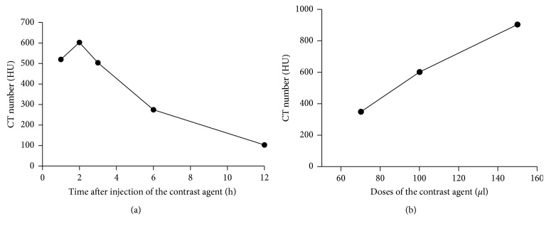
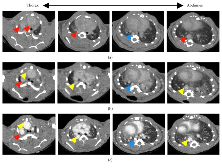
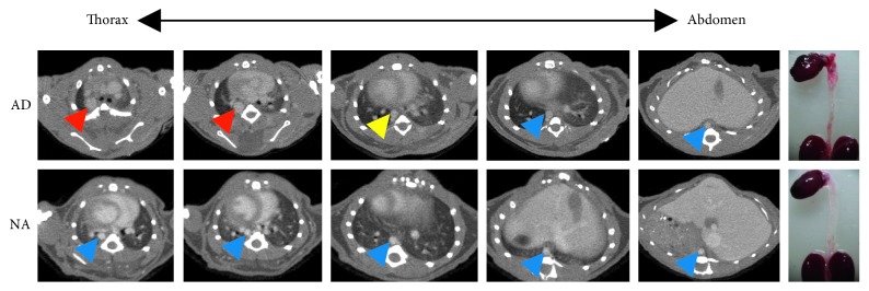
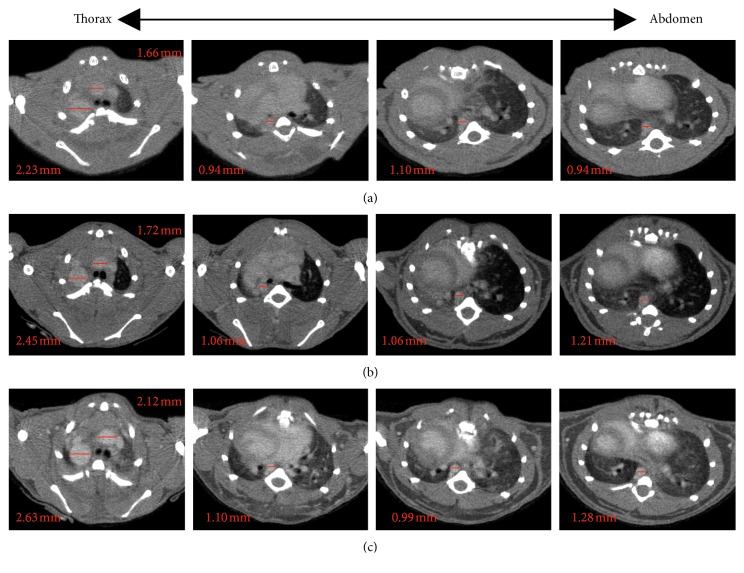
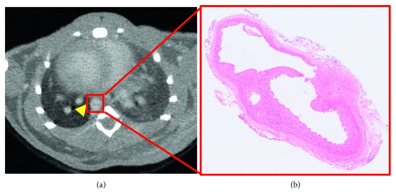
Experiments were performed on healthy mice and mice with aortic dissection. Mice that were developing aortic dissection and healthy mice underwent micro-CT imaging after injection of ExiTron nano 12000. Time-dependent signal enhancement (at 1, 2, 3, 6, and 12 hours after intravenous injection of the contrast agent, respectively) in the aorta of healthy mice was measured to confirm the optimal imaging time of aorta. Various contrast agent doses (70, 100, and 150 l per 25 g mouse, respectively) were investigated to determine the optimal required dose for imaging of the aorta. The mice were scanned with micro-CT at 1, 14, and 28 days after onset of aneurysm and dissection to monitor the dynamic changing process of the aorta. Mouse aortas were stained with hematoxylin and eosin staining, and the diameter of the aorta was measured and compared with those obtained by micro-CT.
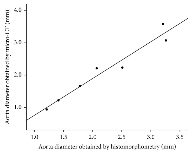
Time-dependent signal enhancement in the aorta shows that the contrast agent has a long blood half-life of 6 hours, with a peak enhancement at 2 hours after injection. Injection of 100 l ExiTron nano 12000 per 25 g mouse allows for effective visualization of the aorta. Micro-CT combined with contrast agent can monitor the changing process of the aorta in the mouse model of aortic aneurysm and dissection dynamically. The values of the diameter of the aortas obtained from the in vivo micro-CT imaging were compared with those obtained from histology and showed a significant correlation ( = 0.96).
These data demonstrate that in vivo micro-CT is an accurate and feasible technique to detect aortic aneurysm or dissection in a mouse model, and the micro-CT technique using the innovative contrast agent ExiTron nano 12000 allows for monitoring various processes dynamically such as aortic remodeling in longitudinal studies.
本研究旨在评估使用血管内对比剂 ExiTron nano 12000 进行血管成像的微计算机断层扫描(micro-CT)的潜力,并监测小鼠主动脉瘤和夹层模型中主动脉的动态变化过程。
在健康小鼠和主动脉夹层模型中进行实验。注射 ExiTron nano 12000 后,对正在发生主动脉夹层的小鼠和健康小鼠进行 micro-CT 成像。测量健康小鼠主动脉的时间依赖性信号增强(分别在静脉注射对比剂后 1、2、3、6 和 12 小时),以确定主动脉的最佳成像时间。研究了不同的对比剂剂量(分别为每 25 克小鼠 70、100 和 150 微升),以确定用于主动脉成像的最佳所需剂量。在动脉瘤和夹层发生后 1、14 和 28 天,用 micro-CT 扫描小鼠,以监测主动脉的动态变化过程。用苏木精和伊红染色法对小鼠主动脉进行染色,并测量主动脉直径,与 micro-CT 获得的直径进行比较。
主动脉的时间依赖性信号增强表明,该对比剂的血液半衰期长,达 6 小时,注射后 2 小时达到峰值增强。每 25 克小鼠注射 100 微升 ExiTron nano 12000 可有效显示主动脉。micro-CT 结合对比剂可动态监测主动脉瘤和夹层模型中主动脉的变化过程。从体内 micro-CT 成像获得的主动脉直径值与组织学获得的直径值之间存在显著相关性(r = 0.96)。
这些数据表明,体内 micro-CT 是一种准确且可行的技术,可用于检测小鼠模型中的主动脉瘤或夹层,使用新型对比剂 ExiTron nano 12000 的 micro-CT 技术可动态监测各种过程,如纵向研究中的主动脉重塑。