Wood Matthew D, Halfpenny Aaron M, Moore Stephen R
OHSU Department of Pathology, Division of Anatomic Pathology, Section of Neuropathology, Oregon Health & Science University, 3181 SW Sam Jackson Park Road, L-113, Portland, OR, 97213, USA.
Knight Diagnostic Laboratories and Department of Molecular and Medical Genetics, Oregon Health & Science University, Portland, OR, 97239, USA.
Diagn Pathol. 2019 Apr 9;14(1):29. doi: 10.1186/s13000-019-0802-8.
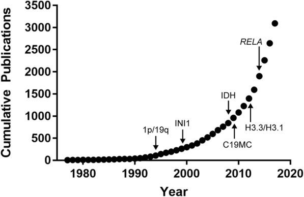
Insights into the molecular underpinnings of primary central nervous system tumors have radically changed the approach to tumor diagnosis and classification. Diagnostic emphasis has shifted from the morphology of a tumor under the microscope to an integrated approach based on morphologic and molecular features, including gene mutations, chromosomal copy number alterations, and gene rearrangements. In 2016, the World Health Organization provided guidelines for making an integrated diagnosis that incorporates both morphologic and molecular features in a subset of brain tumors. The integrated diagnosis now applies to infiltrating gliomas, a category that includes diffusely infiltrating astrocytoma grades II, III, and IV, and oligodendroglioma, grades II and III, thereby encompassing the most common primary intra-axial central nervous system tumors. Other neoplasms such as medulloblastoma, embryonal tumor with multilayered rosettes, certain supratentorial ependymomas, and atypical teratoid/rhabdoid tumor are also eligible for integrated diagnosis, which can sometimes be aided by characteristic immunohistochemical markers. Since 2016, advances in molecular neuro-oncology have resulted in periodic updates and clarifications to the integrated diagnostic approach. These advances reflect expanding knowledge on the molecular pathology of brain tumors, but raise a challenge in rapidly incorporating new molecular findings into diagnostic practice. This review provides a background on the molecular characteristics of primary brain tumors, emphasizing the molecular basis for classification of infiltrating gliomas, the most common entities that are eligible for an integrated diagnosis. We then discuss entities within the diffuse gliomas that do not receive an integrated diagnosis by WHO 2016 criteria, but have distinctive molecular features that are important to recognize because their clinical behavior can influence clinical management and prognosis. Particular attention is given to the histone H3 G34R/G34V mutant astrocytomas, an entity to consider when faced with an infiltrating glioma in the cerebral hemisphere of children and young adults, and to the group of histologically lower grade diffuse astrocytic gliomas with molecular features of glioblastoma, an important category of tumors to recognize due to their aggressive clinical behavior.
对原发性中枢神经系统肿瘤分子基础的深入了解彻底改变了肿瘤诊断和分类的方法。诊断重点已从显微镜下肿瘤的形态学转变为基于形态学和分子特征的综合方法,包括基因突变、染色体拷贝数改变和基因重排。2016年,世界卫生组织提供了在一部分脑肿瘤中结合形态学和分子特征进行综合诊断的指南。综合诊断现在适用于浸润性胶质瘤,这一类别包括弥漫性浸润性II级、III级和IV级星形细胞瘤以及II级和III级少突胶质细胞瘤,从而涵盖了最常见的原发性轴内中枢神经系统肿瘤。其他肿瘤,如髓母细胞瘤、具有多层菊形团的胚胎性肿瘤、某些幕上室管膜瘤和非典型畸胎样/横纹肌样肿瘤也适用于综合诊断,有时特征性免疫组化标志物可辅助诊断。自2016年以来,分子神经肿瘤学的进展导致对综合诊断方法进行了定期更新和阐明。这些进展反映了对脑肿瘤分子病理学认识的不断扩大,但在将新的分子发现迅速纳入诊断实践方面提出了挑战。本综述提供了原发性脑肿瘤分子特征的背景知识,重点强调了浸润性胶质瘤分类的分子基础,浸润性胶质瘤是最常见的适合综合诊断的实体。然后,我们讨论了弥漫性胶质瘤中未根据2016年世界卫生组织标准进行综合诊断,但具有独特分子特征的实体,识别这些特征很重要,因为它们的临床行为会影响临床管理和预后。特别关注组蛋白H3 G34R/G34V突变型星形细胞瘤,在面对儿童和年轻成人脑半球浸润性胶质瘤时需要考虑这一实体;以及具有胶质母细胞瘤分子特征的组织学低级弥漫性星形细胞胶质瘤组,由于其侵袭性临床行为,这是一类需要识别的重要肿瘤。