Yarossi Mathew, Patel Jigna, Qiu Qinyin, Massood Supriya, Fluet Gerard, Merians Alma, Adamovich Sergei, Tunik Eugene
Movement Neuroscience Laboratory, Department of Physical Therapy, Movement and Rehabilitation Science, Bouve College of Health Sciences, Northeastern University, Boston, MA, United States.
SPIRAL Group, Department of Electrical and Computer Engineering, Northeastern University, Boston, MA, United States.
Front Neurol. 2019 Mar 26;10:258. doi: 10.3389/fneur.2019.00258. eCollection 2019.
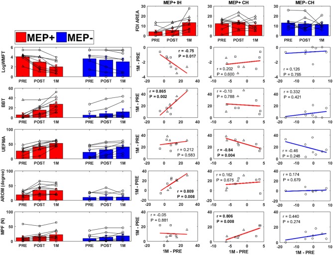
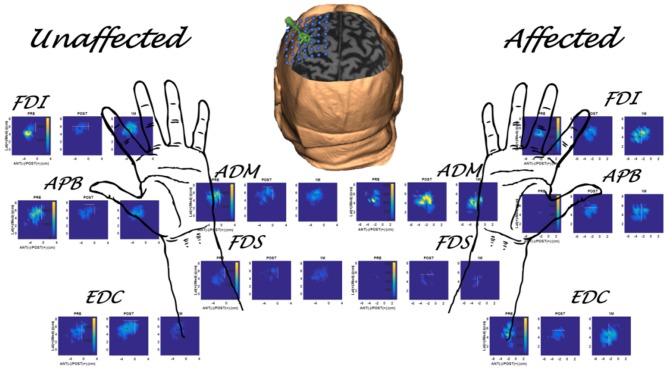
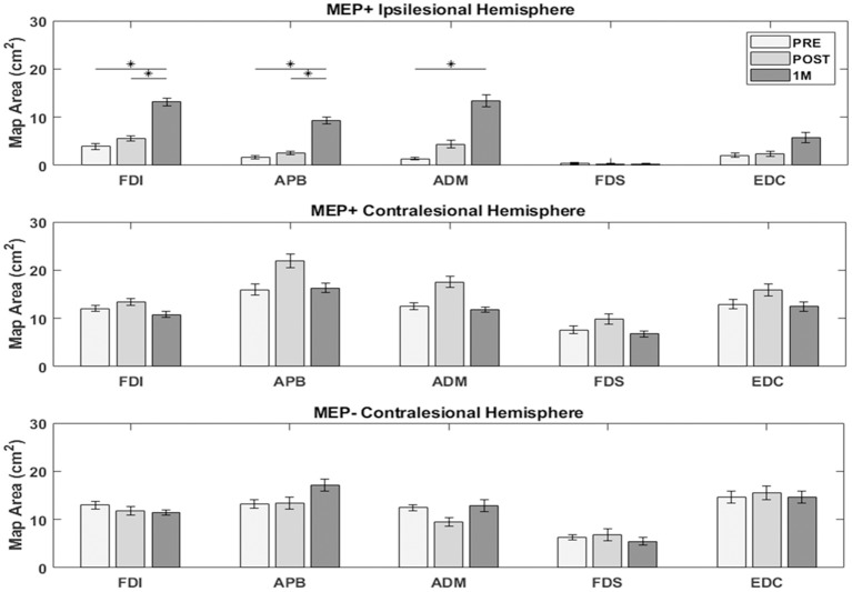
Transcranial magnetic stimulation (TMS) induced motor evoked potentials (MEPs) are an established proxy of corticospinal excitability. As a binary measure, the presence (MEP+) or absence (MEP-) of ipsilesional hemisphere MEPs early following stroke is a robust indicator of long-term recovery, however this measure does not provide information about spatial cortical reorganization. MEPs have been systematically acquired over the sensorimotor cortex to "map" motor topography. In this investigation we compared the degree to which functional improvements resulting from early (<3 months post-stroke) intensive hand focused upper limb rehabilitation correlate with changes in motor topography between MEP+ and MEP- individuals. Following informed consent, 17 individuals (4 Female, 60.3 ± 9.4 years, 24.6 ± 24.01 days post first time stroke) received 8 one hour-sessions of training with virtual reality (VR)/Robotic simulations. Clinical tests [Box and Blocks Test (BBT), Wolf Motor Function Test (WMFT), Upper Extremity Fugl-Meyer (UEFMA)], kinematic and kinetic assessments [finger Active Range of Motion (finger AROM), Maximum Pinch Force (MPF)], and bilateral TMS mapping of 5 hand muscles were performed prior to (PRE), directly following (POST), and 1 month following (1M) training. Participants were divided into two groups (MEP+, MEP-) based on whether an MEP was present in the affected first dorsal interosseous (FDI) at any time point. MEP+ individuals improved significantly more than MEP- individuals from PRE to 1M on the WMFT, BBT, and finger AROM scores. Ipsilesional hemisphere FDI area increased significantly with time in the MEP+ group. FDI area of the contralesional hemisphere was not significantly different across time points or groups. In the MEP+ group, significant correlations were observed between PRE-1M changes in ipsilesional FDI area and WMFT, BBT, and finger AROM, and contralesional FDI area and UEFMA and MPF. In the MEP- group, no significant correlations were found between changes in contralesional FDI area and functional outcomes. We report preliminary evidence in a small sample that patterns of recovery and the association of recovery to bilateral changes in motor topography may depend on integrity of the ipsilesional cortical spinal tract as assessed by the presence of TMS evoked MEPs.
经颅磁刺激(TMS)诱发的运动诱发电位(MEP)是皮质脊髓兴奋性的一种既定替代指标。作为一种二元测量方法,中风后早期患侧半球MEP的存在(MEP+)或缺失(MEP-)是长期恢复的有力指标,然而该测量方法并未提供有关空间皮质重组的信息。已在感觉运动皮层系统地采集MEP以“绘制”运动地形图。在本研究中,我们比较了中风后早期(<3个月)强化手部聚焦上肢康复所带来的功能改善程度与MEP+和MEP-个体之间运动地形图变化的相关性。在获得知情同意后,17名个体(4名女性,60.3±9.4岁,首次中风后24.6±24.01天)接受了8次为时1小时的虚拟现实(VR)/机器人模拟训练。在训练前(PRE)、训练后立即(POST)以及训练后1个月(1M)进行了临床测试[箱块测试(BBT)、沃尔夫运动功能测试(WMFT)、上肢Fugl-Meyer评估(UEFMA)]、运动学和动力学评估[手指主动活动范围(手指AROM)、最大捏力(MPF)]以及对5块手部肌肉进行双侧TMS映射。根据在任何时间点患侧第一背侧骨间肌(FDI)是否存在MEP,将参与者分为两组(MEP+、MEP-)。从PRE到1M,MEP+个体在WMFT、BBT和手指AROM评分上的改善明显大于MEP-个体。MEP+组患侧半球FDI面积随时间显著增加。对侧半球FDI面积在各时间点或组间无显著差异。在MEP+组中,观察到患侧FDI面积从PRE到1M的变化与WMFT、BBT和手指AROM之间存在显著相关性,对侧FDI面积与UEFMA和MPF之间也存在显著相关性。在MEP-组中,未发现对侧FDI面积变化与功能结果之间存在显著相关性。我们在一个小样本中报告了初步证据,即恢复模式以及恢复与运动地形图双侧变化的关联可能取决于通过TMS诱发MEP的存在所评估的患侧皮质脊髓束的完整性。