Nayak Vanishri S, Bhat Nandini, Nayak Sunil S, Sumalatha Suhani
Department of Anatomy, Kasturba Medical College, Manipal Academy of Higher Education, Manipal, India.
Department of Oral and Maxillofacial Surgery, Manipal College of Dental Sciences, Manipal Academy of Higher Education, Manipal, India.
Anat Cell Biol. 2019 Mar;52(1):34-37. doi: 10.5115/acb.2019.52.1.34. Epub 2019 Mar 29.
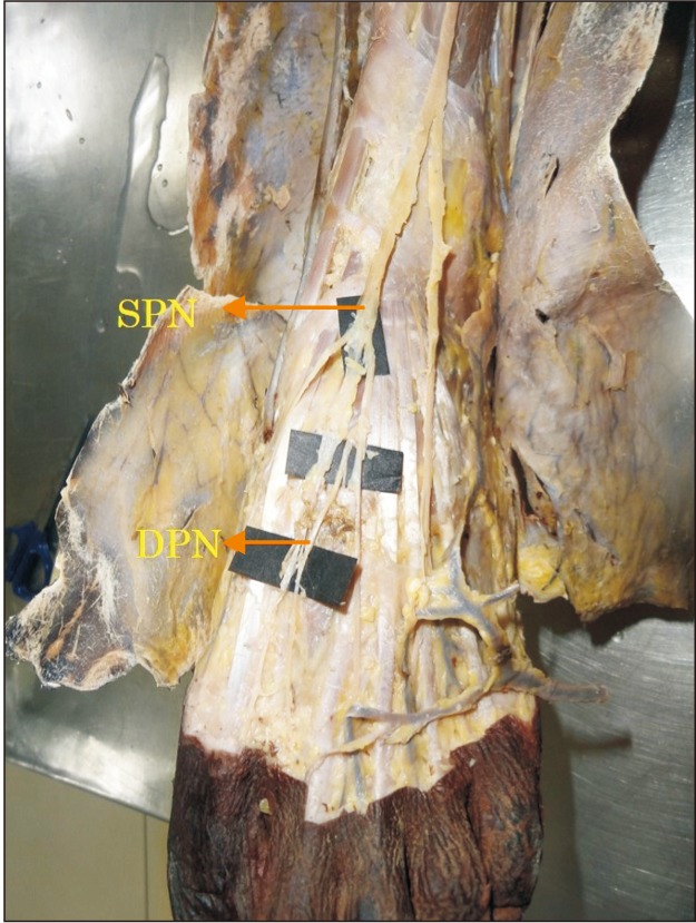
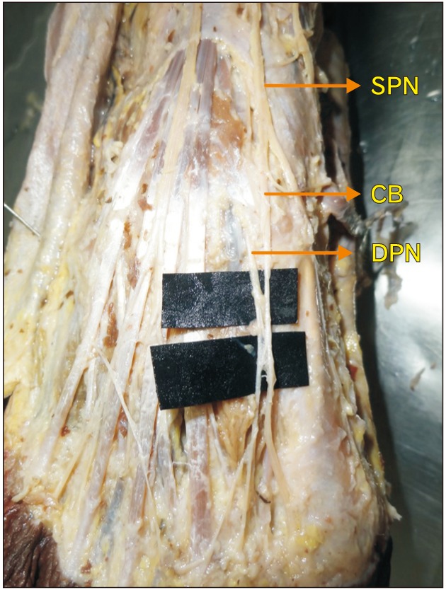
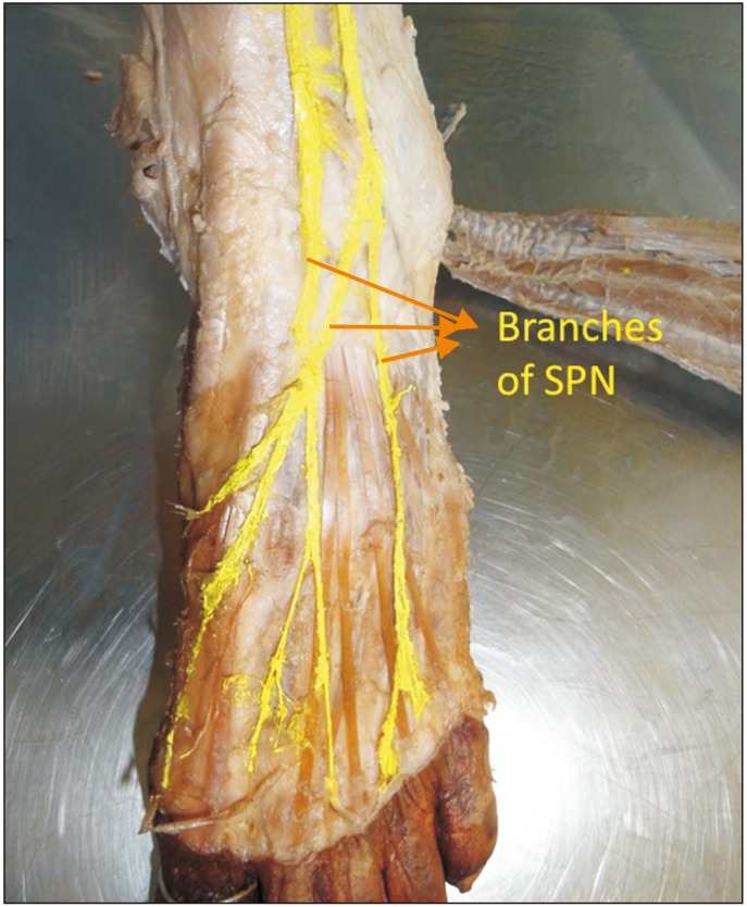
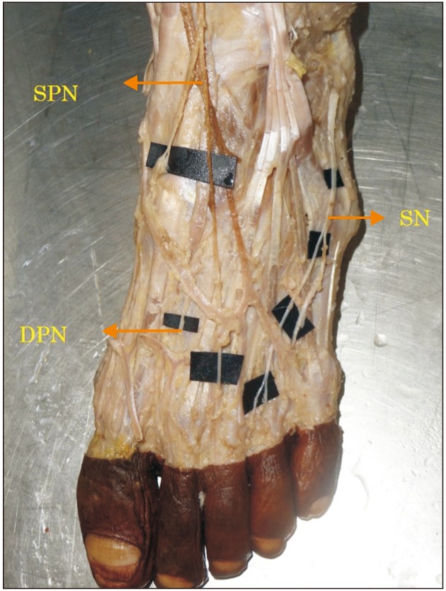
Generally among the branches of common peroneal nerve, the superficial peroneal nerve provides cutaneous innervation to major part of the dorsum of the foot whereas the deep peroneal nerve innervates the skin over the first interdigital cleft region. The sural and saphenous nerves supplies the smaller lateral and medial margins of the dorsum respectively. The present study has been taken to classify the patterns of innervations of the nerves on the dorsum of the foot in South Indian population. A total of 40 formalin fixed lower limbs from 20 adult cadavers (15 males, 5 females) aged between 35 to 60 years were dissected and the branching patterns of nerves on the dorsum of the foot were noted and specimens were photographed. Gross anatomical variations were noted in the branching pattern of superficial peroneal, deep peroneal and sural nerve on the dorsum of foot. Results obtained in our study were classified into four groups. The cutaneous nerves are at risk of iatrogenic injuries during surgeries involving ankle, open reduction and internal fixation of fracture, arthroscopy etc. Knowledge of such anatomical variations of the nerves provides information to clinicians to avoid injury to them in real clinical situations.
一般来说,在腓总神经的分支中,腓浅神经为足背的大部分区域提供皮肤神经支配,而腓深神经支配第一趾蹼间隙区域的皮肤。腓肠神经和隐神经分别供应足背较小的外侧和内侧边缘。本研究旨在对南印度人群足背神经的支配模式进行分类。解剖了20具年龄在35至60岁之间的成年尸体(15例男性,5例女性)的40条福尔马林固定下肢,记录足背神经的分支模式并拍摄标本照片。观察到足背腓浅神经、腓深神经和腓肠神经分支模式的大体解剖变异。我们研究中获得的结果分为四组。在涉及踝关节、骨折切开复位内固定、关节镜检查等手术过程中,皮神经有发生医源性损伤的风险。了解这些神经的解剖变异可为临床医生在实际临床情况中避免损伤它们提供信息。