Asaeda Makoto, Deie Masataka, Kono Yoshifumi, Mikami Yukio, Kimura Hiroaki, Adachi Nobuo
Sports Medical Center, Hiroshima University Hospital, 1-2-3 Kasumi, Minami-ku, Hiroshima, 734-8551, Japan.
Division of Rehabilitation, Department of Clinical Practice and Support, Hiroshima University Hospital, 1-2-3 Kasumi, Minami-ku, Hiroshima, 734-8551, Japan.
Asia Pac J Sports Med Arthrosc Rehabil Technol. 2018 Dec 14;16:14-18. doi: 10.1016/j.asmart.2018.11.004. eCollection 2019 Apr.
Knee joint kinematics and kinetics during running recover at 12 months, not 6 months, following anterior cruciate ligament (ACL) reconstruction surgery. Knee muscle strength is a criterion used to assess an individual's readiness to return-to-sports (RTS); however, the relationship between knee muscle strength and knee biomechanics is unclear. This study investigated the relationship between knee muscle strength and dynamic knee biomechanics during running at 6 and 12 months after ACL reconstruction surgery.
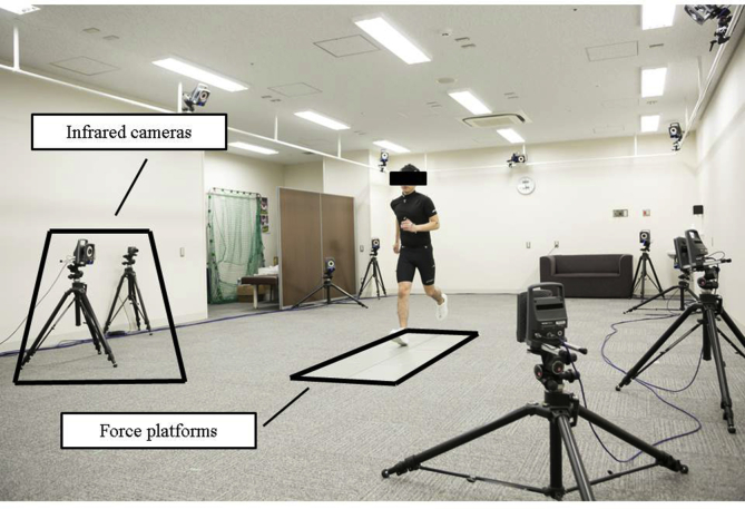
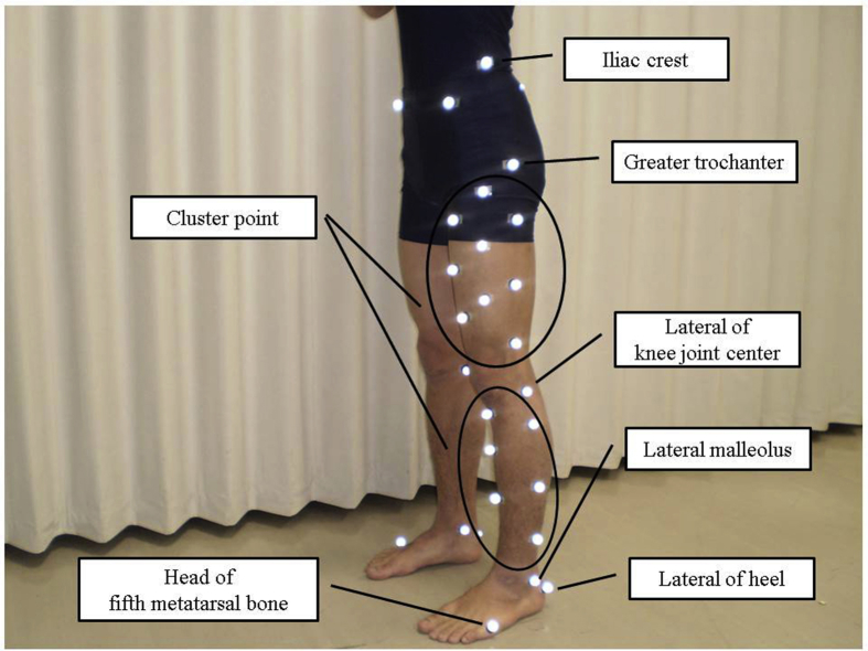
Knee joint kinematics and kinetics during running were analyzed in 21 patients (10 males, 11 females) who underwent ACL reconstruction for a unilateral ACL deficiency. Kinematics and Kinetics were measured by three-dimensional motion analysis system, and Knee flexion angle was calculated using Point cluster technique and internal extension moment was calculated by the inverse dynamics method. Patients were compared to a control group matched by age, height and weight. Isokinetic knee extension and flexion strength in ACL-reconstructed patients were measured at 6 and 12 months postsurgery, by separated gender.
Knee flexion angle was significantly lower in ACL patients at 6 months postsurgery compared to the control group (F (2, 62)=5.78, =0.014). There were significant lower peak knee flexion angles in male groups than female (F (1, 62)=6.33, <0.01). Knee extension moments were significantly lower in both male and female ACL patients compared to the control group at 6 and 12 months postsurgery (F (2, 62)=12.05, <0.01(6 months), =0.034(12 months)), and there were significant correlations with knee extension moments and maximum torque of knee extension/flexion (<0.05). At 12 months after surgery, knee joint kinematics in ACL patients were restored. Both peak knee angle and knee extension moment were significantly associated with maximum knee extension/flexion torque values in female patients at 12 months postsurgery.
Dynamic knee biomechanics during running were not restored 6 and 12 months after ACL reconstruction both male and female. It is necessary to strengthen knee extension and flexion muscles to restore knee kinetics during running, especially female patients.
前交叉韧带(ACL)重建手术后,跑步时的膝关节运动学和动力学在12个月而非6个月时恢复。膝关节肌肉力量是评估个体恢复运动(RTS)准备情况的一个标准;然而,膝关节肌肉力量与膝关节生物力学之间的关系尚不清楚。本研究调查了ACL重建手术后6个月和12个月时跑步过程中膝关节肌肉力量与动态膝关节生物力学之间的关系。
对21例(10例男性,11例女性)因单侧ACL损伤接受ACL重建手术的患者跑步时的膝关节运动学和动力学进行分析。通过三维运动分析系统测量运动学和动力学,使用点簇技术计算膝关节屈曲角度,并通过逆动力学方法计算内翻伸展力矩。将患者与年龄、身高和体重匹配的对照组进行比较。在术后6个月和12个月,按性别分别测量ACL重建患者的等速膝关节伸展和屈曲力量。
与对照组相比,ACL患者术后6个月时膝关节屈曲角度显著更低(F(2, 62)=5.78,P=0.014)。男性组的膝关节屈曲峰值角度显著低于女性(F(1, 62)=6.33,P<0.01)。与对照组相比,男性和女性ACL患者术后6个月和12个月时膝关节伸展力矩均显著更低(F(2, 62)=12.05,P<0.01(6个月),P=0.034(12个月)),并且膝关节伸展力矩与膝关节伸展/屈曲最大扭矩之间存在显著相关性(P<0.05)。术后12个月时,ACL患者的膝关节运动学恢复。术后12个月时,女性患者的膝关节角度峰值和膝关节伸展力矩均与膝关节伸展/屈曲最大扭矩值显著相关。
ACL重建术后6个月和12个月时,男性和女性跑步时的动态膝关节生物力学均未恢复。有必要加强膝关节伸展和屈曲肌肉,以恢复跑步时的膝关节动力学,尤其是女性患者。