Section on Magnetic Spectroscopy, National Institute of Mental Health, National Institutes of Health, Bethesda, Maryland, United States of America.
Functional Magnetic Resonance Imaging Core Facility, National Institute of Mental Health, National Institutes of Health, Bethesda, Maryland, United States of America.
PLoS One. 2019 Apr 17;14(4):e0215210. doi: 10.1371/journal.pone.0215210. eCollection 2019.
The principal excitatory neurotransmitter glutamate plays an important role in many central nervous system disorders. Because glutamate resides predominantly in glutamatergic neurons, its relaxation properties reflect the intracellular environment of glutamatergic neurons. This study developed an improved echo time-independent technique for measuring transverse relaxation time and demonstrated that this radio frequency (RF)-driven longitudinal steady state technique can reliably measure glutamate transverse relaxation in the frontal cortex, where structural and functional abnormalities have been associated with psychiatric symptoms.
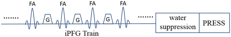
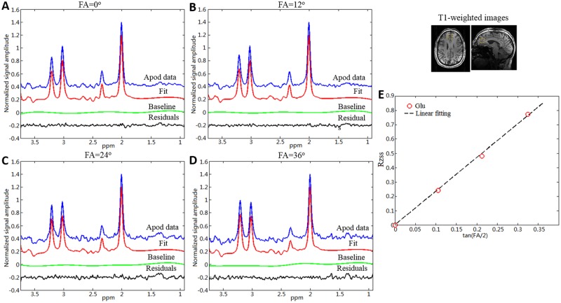
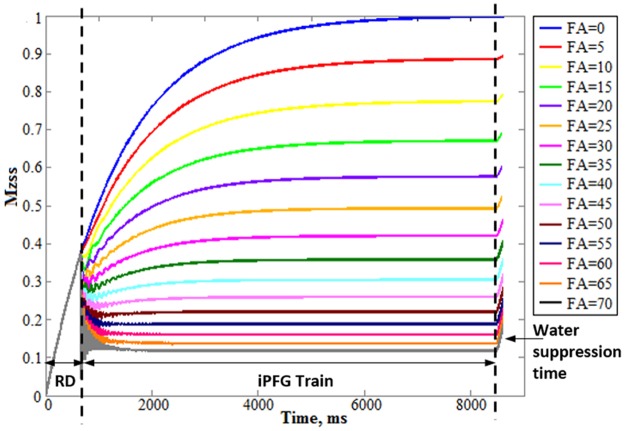
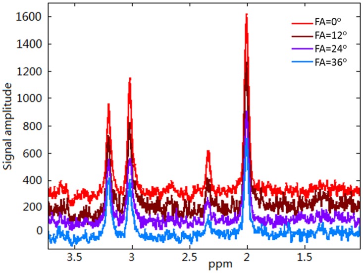
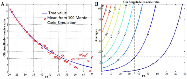
Bloch and Monte Carlo simulations were performed to improve and optimize the RF-driven, longitudinal, steady-state (MARzss) technique to significantly shorten scan time and increase measurement precision. Optimized four-flip angle measurements at 0°,12°, 24°, and 36° with matched repetition time were used in nine human subjects (6F, 3M; 27-49 years old) at 7 Tesla. Longitudinal and transverse relaxation rates for glutamate were measured from a 2 x 2 x 2 cm3 voxel placed in three different brain regions: gray matter-dominated medial prefrontal lobe, white matter-dominated left frontal lobe, and gray matter-dominated occipital lobe.
Compared to the original MARzss technique, the scan time per voxel for measuring glutamate transverse relaxation was shortened by more than 50%. In the medial frontal, left frontal, and occipital voxels, the glutamate T2 was found to be 117.5±12.9 ms (mean ± standard deviation, n = 9), 107.3±12.1 (n = 9), and 124.4±16.6 ms (n = 8), respectively.
The improvements described in this study make the MARZSS technique a viable tool for reliably measuring glutamate relaxation from human subjects in a typical clinical setting. It is expected that this improved technique can be applied to characterize the intracellular environment of glutamatergic neurons in a variety of brain disorders.
主要的兴奋性神经递质谷氨酸在许多中枢神经系统疾病中起着重要作用。由于谷氨酸主要存在于谷氨酸能神经元中,其弛豫特性反映了谷氨酸能神经元的细胞内环境。本研究开发了一种改进的回波时间独立技术来测量横向弛豫时间,并证明这种射频(RF)驱动的纵向稳态技术可以可靠地测量前额皮质中的谷氨酸横向弛豫,其中结构和功能异常与精神症状有关。
进行了布洛赫和蒙特卡罗模拟,以改进和优化射频驱动的、纵向的、稳态(MARzss)技术,以显著缩短扫描时间并提高测量精度。在 7T 下,对 9 名受试者(6 名女性,3 名男性;27-49 岁)进行了优化的四翻转角测量,翻转角为 0°、12°、24°和 36°,匹配重复时间。在三个不同的脑区(灰质为主的内侧前额叶、白质为主的左侧额叶和灰质为主的枕叶)中放置一个 2×2×2cm3 的体素,测量谷氨酸的纵向和横向弛豫率。
与原始 MARzss 技术相比,测量谷氨酸横向弛豫的每个体素的扫描时间缩短了 50%以上。在内侧前额叶、左侧额叶和枕叶体素中,谷氨酸 T2 分别为 117.5±12.9ms(平均值±标准偏差,n=9)、107.3±12.1(n=9)和 124.4±16.6ms(n=8)。
本研究中描述的改进使 MARZss 技术成为一种可靠的工具,可用于在典型的临床环境下从人体受试者中测量谷氨酸弛豫。预计这种改进的技术可以应用于各种脑疾病中描述谷氨酸能神经元的细胞内环境。