• 文献检索
  • 文档翻译
  • 深度研究
  • 学术资讯
  • Suppr Zotero 插件Zotero 插件
  • 邀请有礼
  • 套餐&价格
  • 历史记录
应用&插件
Suppr Zotero 插件Zotero 插件浏览器插件Mac 客户端Windows 客户端微信小程序
定价
高级版会员购买积分包购买API积分包
服务
文献检索文档翻译深度研究API 文档MCP 服务
关于我们
关于 Suppr公司介绍联系我们用户协议隐私条款
关注我们

Suppr 超能文献

核心技术专利:CN118964589B侵权必究
粤ICP备2023148730 号-1Suppr @ 2026

文献检索

告别复杂PubMed语法,用中文像聊天一样搜索,搜遍4000万医学文献。AI智能推荐,让科研检索更轻松。

立即免费搜索

文件翻译

保留排版,准确专业,支持PDF/Word/PPT等文件格式,支持 12+语言互译。

免费翻译文档

深度研究

AI帮你快速写综述,25分钟生成高质量综述,智能提取关键信息,辅助科研写作。

立即免费体验

多动态多回波序列中不同层厚和层间距对 T1 和 T2 值测量的影响。

The Effect of Varying Slice Thickness and Interslice Gap on T and T Measured with the Multidynamic Multiecho Sequence.

机构信息

Department of Radiology, Seoul National University Hospital.

Department of Radiology, Seoul National University College of Medicine.

出版信息

Magn Reson Med Sci. 2019 Apr 10;18(2):126-133. doi: 10.2463/mrms.mp.2018-0010. Epub 2018 Jul 6.

DOI:10.2463/mrms.mp.2018-0010
PMID:29984783
原文链接:https://pmc.ncbi.nlm.nih.gov/articles/PMC6460125/
Abstract

PURPOSE

The purpose of our study was to investigate the effect of different slice thicknesses and/or interslice gaps on longitudinal and transverse relaxation times (T and T) measured by a multi-dynamic, multi-echo (MDME) sequence.

MATERIALS AND METHODS

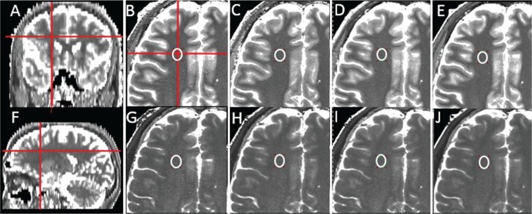
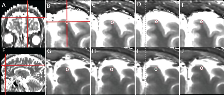
This retrospective study included nine healthy subjects who underwent MDME sequence (at 3T) with four different combinations of slice thicknesses and/or interslice gaps: slice thickness of 4 mm and interslice gap of 0 mm (TH4/G0), TH4/G1, TH5/G0, and TH5/G1. T and T were measured in various brain regions by a qualified neuroradiologist with 8 years of clinical experience: the frontal white matter (WM), occipital WM, genu, splenium, frontal cortex, thalamus, putamen, caudate head, and cerebrospinal fluid (CSF). The paired samples t-test was used to investigate the effect of different slice thicknesses and interslice gaps (TH4/G0 versus TH4/G1 and TH5/G0 versus TH5/G1). P < 0.013 was considered statistically significant.

RESULTS

T in all brain regions and T in the frontal WM, putamen, and CSF did not significantly change for different slice thicknesses and/or gaps (Ps > 0.013). In addition, T in all brain regions of interest did not significantly change between TH4/G0, TH4/G1, TH5/G0 and TH5/G1. However, T in some of the brain regions was higher with TH4/G0 than with TH5/G0 (occipital WM, frontal cortex, and caudate head) and with TH4/G1 than with TH5/G1 (occipital WM, genu, splenium and thalamus, all Ps < 0.013).

CONCLUSION

T estimated using the MDME sequence was stable regardless of slice thickness or gap. Although the sequence seems to provide stable relaxation values, identical slice thicknesses need to be used for follow-up to prevent potential T changes.

摘要

目的

本研究旨在探讨多动态、多回波(MDME)序列测量的不同层厚和/或层间距对纵向弛豫时间(T)和横向弛豫时间(T)的影响。

材料和方法

本回顾性研究纳入了 9 名健康受试者,他们接受了 MDME 序列(3T)检查,共采用了 4 种不同的层厚和/或层间距组合:层厚 4mm 且层间距 0mm(TH4/G0)、TH4/G1、TH5/G0 和 TH5/G1。一位具有 8 年临床经验的合格神经放射科医师在不同脑区测量 T 和 T:额白质(WM)、枕 WM、膝部、压部、额皮质、丘脑、壳核、尾状核头部和脑脊液(CSF)。采用配对样本 t 检验比较不同层厚和层间距(TH4/G0 与 TH4/G1 和 TH5/G0 与 TH5/G1)的影响。P<0.013 认为具有统计学意义。

结果

不同层厚和层间距时,所有脑区的 T 和 T(额 WM、壳核和 CSF)均无显著变化(P>0.013)。此外,TH4/G0、TH4/G1、TH5/G0 和 TH5/G1 之间各感兴趣脑区的 T 也无显著变化。然而,与 TH5/G0 相比,TH4/G0 时一些脑区的 T 更高(枕 WM、额皮质和尾状核头部),TH4/G1 时也更高(枕 WM、膝部、压部和丘脑,所有 P<0.013)。

结论

使用 MDME 序列估计的 T 是稳定的,与层厚或层间距无关。尽管该序列似乎提供了稳定的弛豫值,但为了防止潜在的 T 变化,仍需要使用相同的层厚进行随访。

https://cdn.ncbi.nlm.nih.gov/pmc/blobs/096e/6460125/285fa06ec563/mrms-18-126-g3.jpg
https://cdn.ncbi.nlm.nih.gov/pmc/blobs/096e/6460125/1c78497744bb/mrms-18-126-g1.jpg
https://cdn.ncbi.nlm.nih.gov/pmc/blobs/096e/6460125/0d3714917a9e/mrms-18-126-g2.jpg
https://cdn.ncbi.nlm.nih.gov/pmc/blobs/096e/6460125/285fa06ec563/mrms-18-126-g3.jpg
https://cdn.ncbi.nlm.nih.gov/pmc/blobs/096e/6460125/1c78497744bb/mrms-18-126-g1.jpg
https://cdn.ncbi.nlm.nih.gov/pmc/blobs/096e/6460125/0d3714917a9e/mrms-18-126-g2.jpg
https://cdn.ncbi.nlm.nih.gov/pmc/blobs/096e/6460125/285fa06ec563/mrms-18-126-g3.jpg

相似文献

1
The Effect of Varying Slice Thickness and Interslice Gap on T and T Measured with the Multidynamic Multiecho Sequence.多动态多回波序列中不同层厚和层间距对 T1 和 T2 值测量的影响。
Magn Reson Med Sci. 2019 Apr 10;18(2):126-133. doi: 10.2463/mrms.mp.2018-0010. Epub 2018 Jul 6.
2
Gray matter-white matter contrast on spin-echo T1-weighted images at 3 T and 1.5 T: a quantitative comparison study.3T和1.5T自旋回波T1加权图像上的灰质-白质对比度:一项定量比较研究。
Eur Radiol. 2007 Nov;17(11):2921-5. doi: 10.1007/s00330-007-0688-9. Epub 2007 Jul 7.
3
The effect of scan parameters on T1, T2 relaxation times measured with multi-dynamic multi-echo sequence: a phantom study.多动态多回波序列测量 T1、T2 弛豫时间的扫描参数效应:一项体模研究。
Phys Eng Sci Med. 2022 Jun;45(2):657-664. doi: 10.1007/s13246-022-01128-0. Epub 2022 May 13.
4
Linearity, Bias, Intrascanner Repeatability, and Interscanner Reproducibility of Quantitative Multidynamic Multiecho Sequence for Rapid Simultaneous Relaxometry at 3 T: A Validation Study With a Standardized Phantom and Healthy Controls.3T 下快速同时弛豫率定量多动态多回波序列的线性、偏倚、内扫描仪重复性和扫描仪间可重复性:使用标准化体模和健康对照的验证研究。
Invest Radiol. 2019 Jan;54(1):39-47. doi: 10.1097/RLI.0000000000000510.
5
Multispectral quantitative magnetic resonance imaging of brain iron stores: a theoretical perspective.脑铁储备的多光谱定量磁共振成像:理论视角
Top Magn Reson Imaging. 2006 Feb;17(1):19-30. doi: 10.1097/01.rmr.0000245460.82782.69.
6
Age-Related Changes in Tissue Value Properties in Children: Simultaneous Quantification of Relaxation Times and Proton Density Using Synthetic Magnetic Resonance Imaging.儿童组织值特性的年龄相关性变化:利用合成磁共振成像同时定量弛豫时间和质子密度。
Invest Radiol. 2018 Apr;53(4):236-245. doi: 10.1097/RLI.0000000000000435.
7
Role of T1 mapping to evaluate brain aging in a healthy population.T1 mapping 评估健康人群脑老化的作用。
Clin Imaging. 2020 Jan;59(1):56-60. doi: 10.1016/j.clinimag.2019.09.005. Epub 2019 Nov 11.
8
Three-dimensional high-resolution simultaneous quantitative mapping of the whole brain with 3D-QALAS: An accuracy and repeatability study.三维高分辨率全脑同时定量图谱成像(3D-QALAS):一项准确性和可重复性研究。
Magn Reson Imaging. 2019 Nov;63:235-243. doi: 10.1016/j.mri.2019.08.031. Epub 2019 Aug 21.
9
Magnetic field dependence of the distribution of NMR relaxation times in the living human brain.活体人脑中核磁共振弛豫时间分布的磁场依赖性
MAGMA. 2008 Mar;21(1-2):131-47. doi: 10.1007/s10334-008-0107-5. Epub 2008 Mar 13.
10
Neonatal brain: regional variability of in vivo MR imaging relaxation rates at 3.0 T--initial experience.新生儿脑:3.0T 下活体磁共振成像弛豫率的区域变异性——初步经验
Radiology. 2005 May;235(2):595-603. doi: 10.1148/radiol.2352031769.

引用本文的文献

1
Synthetic MR: Clinical applications in neuroradiology.合成磁共振成像:在神经放射学中的临床应用
Neuroradiology. 2025 Mar;67(3):509-527. doi: 10.1007/s00234-025-03547-8. Epub 2025 Jan 31.
2
Neurodevelopmental outcome in preterm infants with intraventricular hemorrhages: the potential of quantitative brainstem MRI.早产儿脑室出血的神经发育结局:定量脑桥 MRI 的潜力。
Cereb Cortex. 2024 May 2;34(5). doi: 10.1093/cercor/bhae189.
3
Multi-Dynamic-Multi-Echo-based MRI for the Pre-Surgical Determination of Sellar Tumor Consistency: a Quantitative Approach for Predicting Lesion Resectability.

本文引用的文献

1
Synthetic MRI for Clinical Neuroimaging: Results of the Magnetic Resonance Image Compilation (MAGiC) Prospective, Multicenter, Multireader Trial.用于临床神经成像的合成磁共振成像:磁共振图像汇编(MAGiC)前瞻性、多中心、多阅片者试验的结果
AJNR Am J Neuroradiol. 2017 Jun;38(6):1103-1110. doi: 10.3174/ajnr.A5227. Epub 2017 Apr 27.
2
Synthetic MR Imaging in the Diagnosis of Bacterial Meningitis.合成磁共振成像在细菌性脑膜炎诊断中的应用
Magn Reson Med Sci. 2017 Apr 10;16(2):91-92. doi: 10.2463/mrms.ci.2016-0082. Epub 2016 Dec 22.
3
Synthetic MRI in the Detection of Multiple Sclerosis Plaques.
基于多动态多回波的 MRI 在前庭神经鞘瘤术前硬度评估中的应用:一种预测肿瘤可切除性的定量方法。
Clin Neuroradiol. 2024 Sep;34(3):663-673. doi: 10.1007/s00062-024-01407-1. Epub 2024 Apr 19.
4
Quantitative Magnetic Resonance Imaging for Neurodevelopmental Outcome Prediction in Neonates Born Extremely Premature-An Exploratory Study.定量磁共振成像在预测极早产儿神经发育结局中的应用——一项探索性研究。
Clin Neuroradiol. 2024 Jun;34(2):421-429. doi: 10.1007/s00062-023-01378-9. Epub 2024 Jan 30.
5
Application of magnetic resonance image compilation (MAGiC) in the diagnosis of middle-aged and elderly women with osteoporosis.磁共振图像整合(MAGiC)在中老年女性骨质疏松症诊断中的应用。
BMC Med Imaging. 2023 May 15;23(1):63. doi: 10.1186/s12880-023-01010-9.
6
The effect of scan parameters on T1, T2 relaxation times measured with multi-dynamic multi-echo sequence: a phantom study.多动态多回波序列测量 T1、T2 弛豫时间的扫描参数效应:一项体模研究。
Phys Eng Sci Med. 2022 Jun;45(2):657-664. doi: 10.1007/s13246-022-01128-0. Epub 2022 May 13.
7
Different from the Beginning: WM Maturity of Female and Male Extremely Preterm Neonates-A Quantitative MRI Study.与众不同的开端:女性和男性极早产儿 WM 成熟度的定量 MRI 研究。
AJNR Am J Neuroradiol. 2022 Apr;43(4):611-619. doi: 10.3174/ajnr.A7472. Epub 2022 Mar 24.
8
Mapping Human Fetal Brain Maturation In Vivo Using Quantitative MRI.使用定量 MRI 对体内人类胎儿大脑成熟进行映射。
AJNR Am J Neuroradiol. 2021 Nov;42(11):2086-2093. doi: 10.3174/ajnr.A7286. Epub 2021 Sep 9.
9
Impact of Prematurity on the Tissue Properties of the Neonatal Brain Stem: A Quantitative MR Approach.早产儿脑干组织特性的影响:一种定量磁共振方法。
AJNR Am J Neuroradiol. 2021 Mar;42(3):581-589. doi: 10.3174/ajnr.A6945. Epub 2021 Jan 21.
10
Validity of SyMRI for Assessment of the Neonatal Brain.SyMRI 评估新生儿脑的有效性。
Clin Neuroradiol. 2021 Jun;31(2):315-323. doi: 10.1007/s00062-020-00894-2. Epub 2020 Mar 11.
合成磁共振成像在检测多发性硬化斑块中的应用
AJNR Am J Neuroradiol. 2017 Feb;38(2):257-263. doi: 10.3174/ajnr.A5012. Epub 2016 Dec 8.
4
Clinical validation of synthetic brain MRI in children: initial experience.儿童合成脑磁共振成像的临床验证:初步经验
Neuroradiology. 2017 Jan;59(1):43-50. doi: 10.1007/s00234-016-1765-z. Epub 2016 Nov 26.
5
Utility of a Multiparametric Quantitative MRI Model That Assesses Myelin and Edema for Evaluating Plaques, Periplaque White Matter, and Normal-Appearing White Matter in Patients with Multiple Sclerosis: A Feasibility Study.一种评估髓鞘和水肿的多参数定量MRI模型在评估多发性硬化症患者斑块、斑块周围白质和正常外观白质中的效用:一项可行性研究
AJNR Am J Neuroradiol. 2017 Feb;38(2):237-242. doi: 10.3174/ajnr.A4977. Epub 2016 Oct 27.
6
Contrast-enhanced synthetic MRI for the detection of brain metastases.用于检测脑转移瘤的对比增强合成磁共振成像
Acta Radiol Open. 2016 Feb 15;5(2):2058460115626757. doi: 10.1177/2058460115626757. eCollection 2016 Feb.
7
Clinical Feasibility of Synthetic MRI in Multiple Sclerosis: A Diagnostic and Volumetric Validation Study.合成磁共振成像在多发性硬化症中的临床可行性:一项诊断和容积验证研究
AJNR Am J Neuroradiol. 2016 Jun;37(6):1023-9. doi: 10.3174/ajnr.A4665. Epub 2016 Jan 21.
8
Quantitative MRI for Rapid and User-Independent Monitoring of Intracranial CSF Volume in Hydrocephalus.定量磁共振成像用于快速且独立于用户的脑积水颅内脑脊液体积监测。
AJNR Am J Neuroradiol. 2016 May;37(5):797-801. doi: 10.3174/ajnr.A4627. Epub 2015 Dec 24.
9
Accuracy and reproducibility of a quantitative magnetic resonance imaging method for concurrent measurements of tissue relaxation times and proton density.一种用于同时测量组织弛豫时间和质子密度的定量磁共振成像方法的准确性和可重复性。
Magn Reson Imaging. 2015 Jun;33(5):584-91. doi: 10.1016/j.mri.2015.02.013. Epub 2015 Feb 20.
10
Normal appearing and diffusely abnormal white matter in patients with multiple sclerosis assessed with quantitative MR.采用定量磁共振成像评估的多发性硬化症患者中外观正常和弥漫性异常的白质
PLoS One. 2014 Apr 18;9(4):e95161. doi: 10.1371/journal.pone.0095161. eCollection 2014.