Valcz Gábor, Buzás Edit I, Kittel Ágnes, Krenács Tibor, Visnovitz Tamás, Spisák Sándor, Török György, Homolya László, Zsigrai Sára, Kiszler Gábor, Antalffy Géza, Pálóczi Krisztina, Szállási Zoltán, Szabó Vanessza, Sebestyén Anna, Solymosi Norbert, Kalmár Alexandra, Dede Kristóf, Lőrincz Péter, Tulassay Zsolt, Igaz Péter, Molnár Béla
Molecular Medicine Research Group, Hungarian Academy of Sciences and Semmelweis University, Budapest, Hungary.
2nd Department of Medicine, Semmelweis University, Budapest, Hungary.
J Extracell Vesicles. 2019 Apr 8;8(1):1596668. doi: 10.1080/20013078.2019.1596668. eCollection 2019.
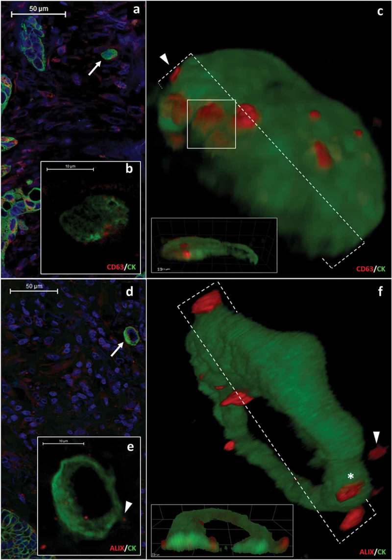
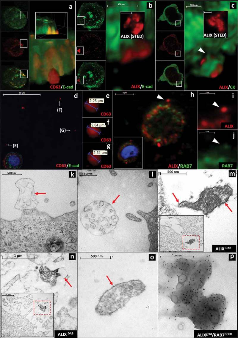
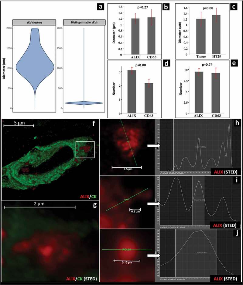
Small extracellular vesicles (EVs) are membrane enclosed structures that are usually released from cells upon exocytosis of multivesicular bodies (MVBs) as a collection of separate, free EVs. In this study, we analysed paraffin embedded sections of archived human colorectal cancer samples. We studied 3D reconstructions of confocal microscopic images complemented by HyVolution and STED imaging. Unexpectedly, we found evidence that large, MVB-like aggregates of ALIX/CD63 positive EV clusters were released by migrating tumour cells. These structures were often captured with partial or complete extra-cytoplasmic localization at the interface of the plasma membrane of the tumour cell and the stroma. Their diameter ranged between 0.62 and 1.94 μm (mean±S.D.: 1.17 ± 0.34 μm). High-resolution 3D reconstruction showed that these extracellular MVB-like EV clusters were composed of distinguishable internal particles of small EV size (mean±S.D.: 128.96 ± 16.73 nm). , HT29 colorectal cancer cells also showed the release of similar structures as confirmed by immunohistochemistry and immune electron microscopy. Our results provide evidence for an transmission of MVB-like EV clusters through the plasma membrane. Immunofluorescent-based detection of the MVB like small EV clusters in archived pathological samples may represent a novel and unique opportunity which enables analysis of EV release in human tissues.
小细胞外囊泡(EVs)是膜包裹的结构,通常在多泡体(MVBs)胞吐时作为单独的游离EVs集合从细胞中释放出来。在本研究中,我们分析了存档的人类结直肠癌样本的石蜡包埋切片。我们研究了通过HyVolution和STED成像补充的共聚焦显微镜图像的三维重建。出乎意料的是,我们发现有证据表明迁移的肿瘤细胞释放出ALIX/CD63阳性EV簇的大的、类似MVB的聚集体。这些结构经常在肿瘤细胞质膜与基质界面处部分或完全位于细胞质外被捕获。它们的直径在0.62至1.94μm之间(平均值±标准差:1.17±0.34μm)。高分辨率三维重建显示,这些细胞外类似MVB的EV簇由小EV大小的可区分内部颗粒组成(平均值±标准差:128.96±16.73nm)。免疫组织化学和免疫电子显微镜证实,HT29结肠癌细胞也显示出类似结构的释放。我们的结果为类似MVB的EV簇通过质膜的传递提供了证据。基于免疫荧光检测存档病理样本中类似MVB的小EV簇可能代表了一个新的独特机会,能够分析人类组织中的EV释放情况。