Department of Radiology, Isala, Dokter van Heesweg 2, 8025 AB, Zwolle, The Netherlands.
Department of Radiology and Nuclear Medicine, Amsterdam UMC, University of Amsterdam, Amsterdam Movement Sciences, Amsterdam, The Netherlands.
Skeletal Radiol. 2019 Nov;48(11):1775-1785. doi: 10.1007/s00256-019-03206-z. Epub 2019 Apr 24.
To evaluate the impact of radiation dose reduction on image quality in patients with metal-on-metal total hip arthroplasties (THAs) using model-based iterative reconstruction (MBIR) combined with orthopaedic metal artefact reduction (O-MAR).
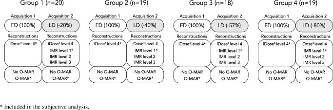
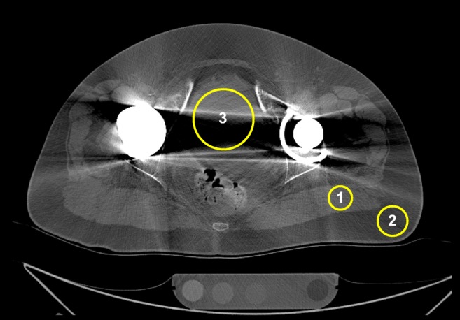
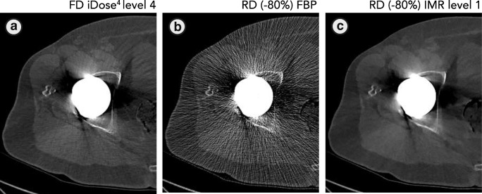
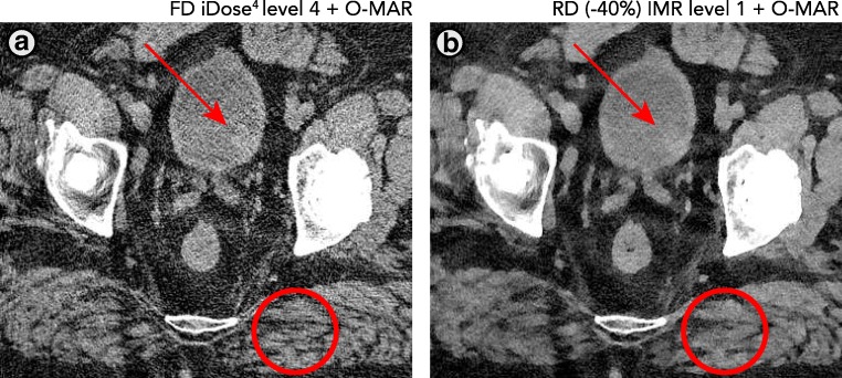
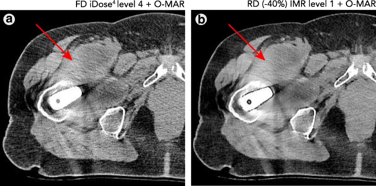
Patients with metal-on-metal THAs received a pelvic CT with a full (FD) and a reduced radiation dose (RD) with -20%, -40%, -57%, or -80% CT radiation dose respectively, when assigned to group 1, 2, 3, or 4 respectively. FD acquisitions were reconstructed with iterative reconstruction, iDose. RD acquisitions were additionally reconstructed with iterative model-based reconstruction (IMR) levels 1-3 with different levels of noise suppression. CT numbers, noise and contrast-to-noise ratios were measured in muscle, fat and bladder. Subjective image quality was evaluated on seven aspects including artefacts, osseous structures, prosthetic components and soft tissues.
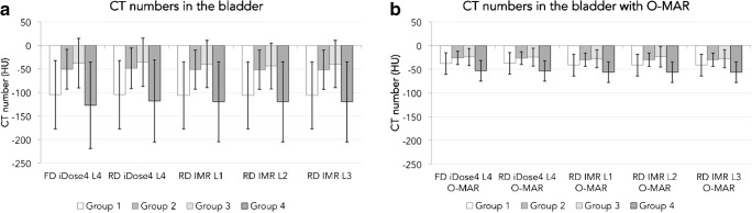
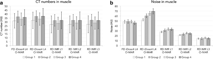
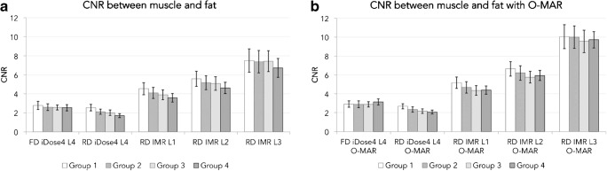
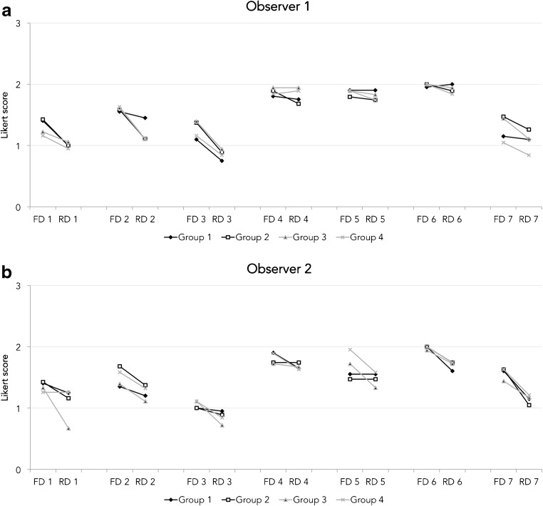
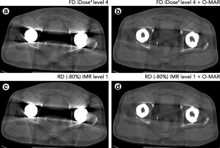
Seventy-six patients were randomly assigned to one of the four groups. While reducing radiation dose by 20%, 40%, 57%, or 80% in combination with IMR, CT numbers remained constant. Compared with iDose, the noise decreased (p < 0.001) and contrast-to-noise ratios increased (p < 0.001) with IMR. O-MAR improved CT number accuracy in the bladder and reduced noise in the bladder, muscle and fat (p < 0.01). Subjective image quality was rated lower on RD IMR images than FD iDose images on all seven aspects (p < 0.05) and was not related to the applied radiation dose reduction.
In RD IMR with O-MAR images, CT numbers remained constant, noise decreased and contrast-to-noise ratios between muscle and fat increased compared with FD iDose with O-MAR images in patients with metal-on-metal THAs. Subjective image quality reduced, regardless of the degree of radiation dose reduction.
使用基于模型的迭代重建(MBIR)结合矫形金属伪影减少(O-MAR)技术,评估金属对金属全髋关节置换术(THA)患者减少放射剂量对图像质量的影响。
将金属对金属 THA 患者随机分为 4 组,每组分别接受全剂量(FD)和降低 20%、40%、57%或 80%的辐射剂量(RD)的骨盆 CT 检查。FD 采集用迭代重建 iDose 重建,RD 采集用迭代模型重建(IMR)1-3 级,并采用不同水平的噪声抑制。在肌肉、脂肪和膀胱中测量 CT 值、噪声和对比噪声比。从 7 个方面评估图像质量,包括伪影、骨结构、假体部件和软组织。
76 例患者随机分为 4 组中的 1 组。与 iDose 相比,当放射剂量分别降低 20%、40%、57%和 80%时,结合 IMR 技术 CT 值保持不变。与 iDose 相比,IMR 降低了噪声(p<0.001),提高了对比噪声比(p<0.001)。O-MAR 提高了膀胱 CT 值的准确性,降低了膀胱、肌肉和脂肪的噪声(p<0.01)。与 FD iDose 图像相比,RD IMR 图像的所有 7 个方面的主观图像质量评分均较低(p<0.05),与应用的放射剂量降低无关。
在 RD IMR 结合 O-MAR 图像中,与 FD iDose 结合 O-MAR 图像相比,金属对金属 THA 患者的 CT 值保持不变,噪声降低,肌肉和脂肪之间的对比噪声比增加。无论放射剂量降低程度如何,主观图像质量均降低。