• 文献检索
  • 文档翻译
  • 深度研究
  • 学术资讯
  • Suppr Zotero 插件Zotero 插件
  • 邀请有礼
  • 套餐&价格
  • 历史记录
应用&插件
Suppr Zotero 插件Zotero 插件浏览器插件Mac 客户端Windows 客户端微信小程序
定价
高级版会员购买积分包购买API积分包
服务
文献检索文档翻译深度研究API 文档MCP 服务
关于我们
关于 Suppr公司介绍联系我们用户协议隐私条款
关注我们

Suppr 超能文献

核心技术专利:CN118964589B侵权必究
粤ICP备2023148730 号-1Suppr @ 2026

文献检索

告别复杂PubMed语法,用中文像聊天一样搜索,搜遍4000万医学文献。AI智能推荐,让科研检索更轻松。

立即免费搜索

文件翻译

保留排版,准确专业,支持PDF/Word/PPT等文件格式,支持 12+语言互译。

免费翻译文档

深度研究

AI帮你快速写综述,25分钟生成高质量综述,智能提取关键信息,辅助科研写作。

立即免费体验

颞下颌关节解剖结构的变化、髁突移位的变化及其与盘移位的关系:磁共振成像研究

Changes in temporomandibular joint anatomy, changes in condylar translation, and their relationship with disc displacement: magnetic resonance imaging study.

作者信息

Bedran Luciane Marie, Dos Santos Alair Augusto Sarmet Moreira Damas

机构信息

Universidade Federal Fluminense (UFF), Niterói, RJ, Brazil.

出版信息

Radiol Bras. 2019 Mar-Apr;52(2):85-91. doi: 10.1590/0100-3984.2018.0020.

DOI:10.1590/0100-3984.2018.0020
PMID:31019336
原文链接:https://pmc.ncbi.nlm.nih.gov/articles/PMC6472865/
Abstract

OBJECTIVE

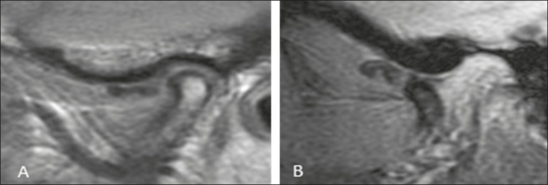
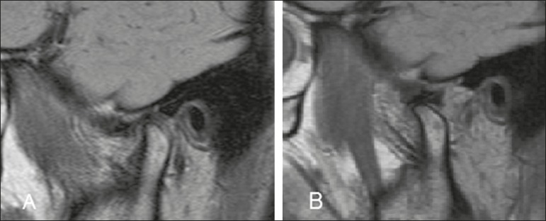
To assess changes in the articular surfaces of the temporomandibular joint (TMJ) and in condylar translation, as detected by magnetic resonance imaging (MRI), determining whether such changes correlate with disc displacement.

MATERIALS AND METHODS

We retrospectively analyzed the MRI scans of 2076 TMJs of 1038 patients with symptoms of temporomandibular disorder. We attempted to determine whether articular disc deformity and changes in condylar translation, as well as changes in the articular surfaces of the condyle, glenoid fossa, and articular eminence, correlated with disc displacement.

RESULTS

Disc displacement with reduction was associated with changes in the shape of the articular eminence. Disc displacement without reduction was most strongly associated with disc deformity, condylar degeneration, glenoid fossa degeneration, and effusion. Neither decreases nor increases in condylar translation were associated with disc deformity, degenerative bone changes, or disc displacement.

CONCLUSION

Changes in the shape of the articular eminence seem to predispose to progression of internal derangement of the TMJ.

摘要

目的

通过磁共振成像(MRI)评估颞下颌关节(TMJ)关节面及髁突移位的变化,确定这些变化是否与盘移位相关。

材料与方法

我们回顾性分析了1038例有颞下颌关节紊乱症状患者的2076个TMJ的MRI扫描图像。我们试图确定关节盘畸形、髁突移位变化以及髁突、关节窝和关节结节关节面的变化是否与盘移位相关。

结果

可复性盘移位与关节结节形状改变有关。不可复性盘移位与盘畸形、髁突退变、关节窝退变及积液关系最为密切。髁突移位的减少或增加均与盘畸形、骨质退行性改变或盘移位无关。

结论

关节结节形状的改变似乎易导致TMJ内紊乱的进展。

https://cdn.ncbi.nlm.nih.gov/pmc/blobs/a51f/6472865/97c44e077ccb/rb-52-02-0085-g03.jpg
https://cdn.ncbi.nlm.nih.gov/pmc/blobs/a51f/6472865/506c81cc6be2/rb-52-02-0085-g01.jpg
https://cdn.ncbi.nlm.nih.gov/pmc/blobs/a51f/6472865/4506bba4c9c6/rb-52-02-0085-g02.jpg
https://cdn.ncbi.nlm.nih.gov/pmc/blobs/a51f/6472865/97c44e077ccb/rb-52-02-0085-g03.jpg
https://cdn.ncbi.nlm.nih.gov/pmc/blobs/a51f/6472865/506c81cc6be2/rb-52-02-0085-g01.jpg
https://cdn.ncbi.nlm.nih.gov/pmc/blobs/a51f/6472865/4506bba4c9c6/rb-52-02-0085-g02.jpg
https://cdn.ncbi.nlm.nih.gov/pmc/blobs/a51f/6472865/97c44e077ccb/rb-52-02-0085-g03.jpg

相似文献

1
Changes in temporomandibular joint anatomy, changes in condylar translation, and their relationship with disc displacement: magnetic resonance imaging study.颞下颌关节解剖结构的变化、髁突移位的变化及其与盘移位的关系:磁共振成像研究
Radiol Bras. 2019 Mar-Apr;52(2):85-91. doi: 10.1590/0100-3984.2018.0020.
2
MRI investigation of TMJ disc and articular eminence morphology in patients with disc displacement.MRI 对颞下颌关节盘和关节突形态的研究在盘移位患者中的应用。
J Stomatol Oral Maxillofac Surg. 2021 Feb;122(1):3-6. doi: 10.1016/j.jormas.2020.09.019. Epub 2020 Oct 12.
3
Disk displacement, eccentric condylar position, osteoarthrosis - misnomers for variations of normality? Results and interpretations from an MRI study in two age cohorts.盘状移位、髁突偏心位置、骨关节炎——正常变异的误称?两个年龄队列MRI研究的结果与解读
BMC Oral Health. 2016 Nov 17;16(1):124. doi: 10.1186/s12903-016-0319-4.
4
Correlation between MRI evidence of degenerative condylar surface changes, induction of articular disc displacement and pathological joint sounds in the temporomandibular joint.颞下颌关节髁突表面退变改变的MRI证据、关节盘移位的诱发与病理性关节弹响之间的相关性。
Gerodontology. 2008 Dec;25(4):251-7. doi: 10.1111/j.1741-2358.2008.00219.x. Epub 2008 Feb 27.
5
[Morphometric analysis of the temporomandibular joint with MRI in 320 joints].[320个颞下颌关节的MRI形态计量分析]
Rofo. 2005 Feb;177(2):217-28. doi: 10.1055/s-2004-813871.
6
Magnetic resonance imaging findings and clinical symptoms in the temporomandibular joint in patients with mandibular continuity defects.下颌连续性缺损患者颞下颌关节的磁共振成像表现及临床症状
J Oral Maxillofac Surg. 2000 May;58(5):487-93; discussion 493-4. doi: 10.1016/s0278-2391(00)90007-7.
7
Association between bone morphology and sagittal disc position in temporomandibular joints of patients with anterior disc displacement.前盘移位患者颞下颌关节的骨形态与矢状位盘位置之间的关联
Hua Xi Kou Qiang Yi Xue Za Zhi. 2022 Jul 25;40(4):414-421. doi: 10.7518/hxkq.2022.04.007.
8
MRI analysis of the relationship between bone changes in the temporomandibular joint and articular disc position in symptomatic patients.MRI 分析症状患者颞下颌关节骨变化与关节盘位置的关系。
Dentomaxillofac Radiol. 2012 Jul;41(5):367-72. doi: 10.1259/dmfr/79317853. Epub 2012 Jan 12.
9
Analysis of the relationship between condylar changes and anterior disc displacement with reduction: a preliminary study.髁突变化与可复性盘前移位关系的分析:一项初步研究。
Oral Radiol. 2023 Jan;39(1):154-163. doi: 10.1007/s11282-022-00617-y. Epub 2022 May 13.
10
Correlation between direction and severity of temporomandibular joint disc displacement and reduction ability during mouth opening.颞下颌关节盘移位方向与严重程度和张口时复位能力之间的相关性
J Oral Rehabil. 2017 Dec;44(12):957-963. doi: 10.1111/joor.12576. Epub 2017 Oct 6.

引用本文的文献

1
Classification of the temporomandibular joint disc displacement without reduction using MRI in the mouth-opening position.使用MRI在开口位对不可复性颞下颌关节盘移位进行分类。
Clin Oral Investig. 2025 May 31;29(6):322. doi: 10.1007/s00784-025-06408-z.
2
A Bibliometric Analysis of Research on Temporomandibular Joint Disc Displacement from 1992 to 2022.1992年至2022年颞下颌关节盘移位研究的文献计量分析
Healthcare (Basel). 2023 Jul 24;11(14):2108. doi: 10.3390/healthcare11142108.
3
A Semi-Automatic Approach for Holistic 3D Assessment of Temporomandibular Joint Changes.

本文引用的文献

1
Value of ultrasound in the anatomical evaluation of the brachial plexus: correlation with magnetic resonance imaging.超声在臂丛神经解剖学评估中的价值:与磁共振成像的相关性
Radiol Bras. 2018 Nov-Dec;51(6):358-365. doi: 10.1590/0100-3984.2017.0083.
2
Contribution of dynamic contrast enhancement and diffusion-weighted magnetic resonance imaging to the diagnosis of malignant cervical lymph nodes.动态对比增强和扩散加权磁共振成像对恶性颈部淋巴结诊断的贡献。
Radiol Bras. 2018 May-Jun;51(3):IX. doi: 10.1590/0100-3984.2018.51.3e3.
3
Magnetic resonance imaging: dynamic contrast enhancement and diffusion-weighted imaging to identify malignant cervical lymph nodes.
一种用于颞下颌关节变化整体三维评估的半自动方法。
J Pers Med. 2023 Feb 16;13(2):343. doi: 10.3390/jpm13020343.
4
Action of Hyaluronic Acid as a Damage-Associated Molecular Pattern Molecule and Its Function on the Treatment of Temporomandibular Disorders.透明质酸作为损伤相关分子模式分子的作用及其在颞下颌关节紊乱病治疗中的功能
Front Pain Res (Lausanne). 2022 Mar 18;3:852249. doi: 10.3389/fpain.2022.852249. eCollection 2022.
5
Cone-beam computed tomographic assessment of the inclination of the articular eminence in patients with temporomandibular disorders and chewing side preference.颞下颌关节紊乱病患者和咀嚼侧偏侧患者关节结节倾斜度的锥形束 CT 评估。
BMC Oral Health. 2021 Aug 13;21(1):396. doi: 10.1186/s12903-021-01760-4.
6
Automated cortical thickness measurement of the mandibular condyle head on CBCT images using a deep learning method.使用深度学习方法在锥形束计算机断层扫描(CBCT)图像上自动测量下颌髁突头部的皮质厚度。
Sci Rep. 2021 Jul 21;11(1):14852. doi: 10.1038/s41598-021-94362-7.
7
Temporomandibular Joints' Morphology and Osteoarthritic Changes in Cone-Beam Computed Tomography Images in Patients with and without Reciprocal Clicking-A Case Control Study.颞下颌关节形态及在伴有和不伴有弹响的患者锥形束 CT 图像中的骨关节炎改变:一项病例对照研究。
Int J Environ Res Public Health. 2020 May 14;17(10):3428. doi: 10.3390/ijerph17103428.
8
Evaluation of temporomandibular disorders by magnetic resonance imaging.通过磁共振成像评估颞下颌关节紊乱病
Radiol Bras. 2019 May-Jun;52(3):VII-VIII. doi: 10.1590/0100-3984.2019.52.3e2.
磁共振成像:动态对比增强及扩散加权成像用于识别恶性颈部淋巴结
Radiol Bras. 2018 Mar-Apr;51(2):71-75. doi: 10.1590/0100-3984.2017.0005.
4
Squamous cell carcinoma of the paranasal sinuses: cutaneous metastases with bone involvement.鼻窦鳞状细胞癌:伴骨受累的皮肤转移
Radiol Bras. 2018 Jan-Feb;51(1):61-62. doi: 10.1590/0100-3984.2016.0113.
5
Perineural spread of malignant head and neck tumors: review of the literature and analysis of cases treated at a teaching hospital.头颈部恶性肿瘤的神经周围扩散:文献综述及教学医院治疗病例分析
Radiol Bras. 2017 Sep-Oct;50(5):323-327. doi: 10.1590/0100-3984.2015.0215.
6
Condyle Excursion Angle, Articular Eminence Inclination, and Temporomandibular Joint Morphologic Relations With Disc Displacement.髁突偏移角度、关节结节斜度以及颞下颌关节形态与盘移位的关系
J Oral Maxillofac Surg. 2017 May;75(5):938.e1-938.e10. doi: 10.1016/j.joms.2017.01.019. Epub 2017 Jan 29.
7
Is Mandibular Fossa Morphology and Articular Eminence Inclination Associated with Temporomandibular Dysfunction?下颌窝形态和关节结节倾斜度与颞下颌关节紊乱病有关联吗?
J Dent (Shiraz). 2016 Jun;17(2):134-41.
8
The value of MRI in patients with temporomandibular joint dysfunction: Correlation of MRI and clinical findings.磁共振成像在颞下颌关节紊乱病患者中的价值:磁共振成像与临床检查结果的相关性
Eur J Radiol. 2016 Apr;85(4):714-9. doi: 10.1016/j.ejrad.2016.02.001. Epub 2016 Feb 4.
9
Non-joint effusion is associated with osteoarthritis in temporomandibular joints with disk displacement.非关节积液与伴有盘移位的颞下颌关节骨关节炎相关。
J Craniomaxillofac Surg. 2016 Jan;44(1):1-5. doi: 10.1016/j.jcms.2015.06.038. Epub 2015 Jul 8.
10
Temporomandibular Joint Disorders and Orofacial Pain.颞下颌关节紊乱病与口面部疼痛
Dent Clin North Am. 2016 Jan;60(1):105-24. doi: 10.1016/j.cden.2015.08.004. Epub 2015 Oct 21.