Costa Lucas E S, Clementino-Neto José, Mendes Carmelita B, Franzon Nayara H, Costa Eduardo de Oliveira, Moura-Neto Vivaldo, Ximenes-da-Silva Adriana
Instituto de Ciências Biológicas e da Saúde, Universidade Federal de Alagoas, Maceió, Brazil.
Instituto do Cérebro and Universidade Federal do Rio de Janeiro, Rio de Janeiro, Brazil.
Front Neurosci. 2019 Apr 4;13:317. doi: 10.3389/fnins.2019.00317. eCollection 2019.
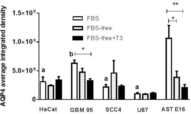
Accumulating evidence indicates that thyroid function and the thyroid hormones L-thyroxine (T4) and L-triiodothyronine (T3) are important factors contributing to the improvement of various pathologies of the central nervous system, including stroke, and various types of cancer, including glioblastoma multiforme (GBM). Low levels of T3 are correlated with the poorest outcome of post-stroke brain function, as well as an increased migration and proliferation of GBM tumor cells. Thyroid hormones are known to stimulate maturation and brain development. Aquaporin 4 (AQP4) is a key factor mediating the cell swelling and edema that occurs during ischemic stroke, and plays a potential role in the migration and proliferation of GBM tumor cells. In this study, as a possible therapeutic target for GBM, we investigated the potential role of T3 in the expression of AQP4 during different stages of mouse brain development. Pregnant mice at gestational day 18, or young animals at postnatal days 27 and 57, received injection of T3 (1 μg/g) or NaOH (0.02 N vehicle). The brains of mice sacrificed on postnatal days 0, 30, and 60 were perfused with 4% paraformaldehyde and sections were prepared for immunohistochemistry of AQP4. AQP4 immunofluorescence was measured in the mouse brains and human GBM cell lines. We found that distribution of AQP4 was localized in astrocytes of the periventricular, subpial, and cerebral parenchyma. Newborn mice treated with T3 showed a significant decrease in AQP4 immunoreactivity followed by an increased expression at P30 and a subsequent stabilization of aquaporin levels in adulthood. All GBM cell lines examined exhibited significantly lower AQP4 expression than cultured astrocytes. T3 treatment significantly downregulated AQP4 in GBM-95 cells but did not influence the rate of GBM cell migration measured 24 h after treatment initiation. Collectively, our results showed that AQP4 expression is developmentally regulated by T3 in astrocytes of the cerebral cortex of newborn and young mice, and is discretely downregulated in GBM cells. These findings indicate that higher concentrations of T3 thyroid hormone would be more suitable for reducing AQP4 in GBM tumorigenic cells, thereby resulting in better outcomes regarding the reduction of brain tumor cell migration and proliferation.
越来越多的证据表明,甲状腺功能以及甲状腺激素左旋甲状腺素(T4)和左旋三碘甲状腺原氨酸(T3)是促进包括中风在内的中枢神经系统各种病理状况改善以及包括多形性胶质母细胞瘤(GBM)在内的各种类型癌症改善的重要因素。低水平的T3与中风后脑功能的最差预后以及GBM肿瘤细胞迁移和增殖增加相关。已知甲状腺激素可刺激成熟和大脑发育。水通道蛋白4(AQP4)是介导缺血性中风期间发生的细胞肿胀和水肿的关键因素,并且在GBM肿瘤细胞的迁移和增殖中发挥潜在作用。在本研究中,作为GBM可能的治疗靶点,我们研究了T3在小鼠脑发育不同阶段对AQP4表达的潜在作用。妊娠第18天的怀孕小鼠或出生后第27天和第57天的幼龄动物接受T3(1μg/g)或NaOH(0.02N载体)注射。在出生后第0、30和60天处死的小鼠的大脑用4%多聚甲醛灌注,并制备切片用于AQP4的免疫组织化学。在小鼠脑和人GBM细胞系中测量AQP4免疫荧光。我们发现AQP4的分布定位于脑室周围、软脑膜下和脑实质的星形胶质细胞中。用T3处理的新生小鼠显示AQP4免疫反应性显著降低,随后在P30时表达增加,成年后水通道蛋白水平随后稳定。所有检测的GBM细胞系均显示AQP4表达明显低于培养的星形胶质细胞。T3处理显著下调GBM-95细胞中的AQP4,但不影响处理开始后24小时测量的GBM细胞迁移率。总体而言,我们的结果表明,AQP4表达在新生和幼龄小鼠大脑皮质的星形胶质细胞中受T3的发育调控,并且在GBM细胞中被离散下调。这些发现表明,更高浓度的T3甲状腺激素更适合降低GBM致瘤细胞中的AQP4,从而在减少脑肿瘤细胞迁移和增殖方面产生更好的结果。