Department of Neurosurgery, St. Peter's Hospital, Seoul 135-809, Republic of Korea.
Biomed Res Int. 2019 Mar 24;2019:6078469. doi: 10.1155/2019/6078469. eCollection 2019.
The purpose of our study is to compare the results of spinal decompression using the full-endoscopic interlaminar technique, tubular retractor, and a conventional microsurgical laminotomy technique and evaluate the advantages and clinical feasibility of minimally invasive spinal (MIS) lumbar decompression technique in the lumbar canal and lateral recess stenosis.
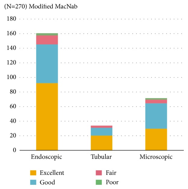
The authors retrospectively reviewed clinical and radiological data from 270 patients who received microsurgical (group E: 72 patients), tubular (group T: 34 patients), or full-endoscopic decompression surgery (group E: 164 patients) for their lumbar canal and lateral recess stenosis from June 2016 to August 2017. Clinical (VAS, ODI, and Mcnab criteria), radiologic (spinal canal diameter, segmental dynamic angle, and disc height), and surgical outcome parameters (CPK level, Operative time, blood loss, and hospital stay) were evaluated pre- and postoperatively and compared among the three groups by means of statistical analysis. Failed cases and complications were reviewed in all groups.
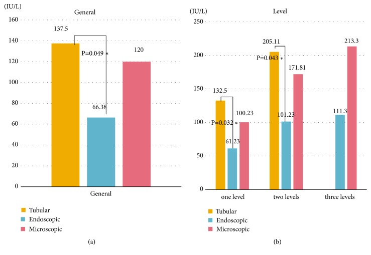
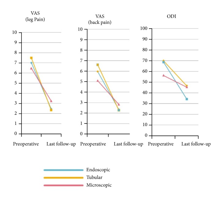
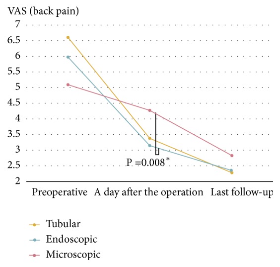
The mean follow-up period was 6.38 months. The Overall clinical success rate was 89.4%. All groups showed favorable clinical outcome. The clinical and radiologic results were similar in all groups. Regarding surgical outcome, group E showed longer operation time than group M and T (group E: 84.17 minutes/level, group M: 52.22 minutes/level, and group T: 66.12 minutes/level) (p<0.05). However, groups E and T showed minimal surgical invasiveness compared with group M. Groups E and T showed less immediate postoperative back pain (VAS) (group E: 3.13, group M: 4.28, group T: 3.54) (p<0.05), less increase of serum CPK enzyme (group E: 66.38 IU/L, group M: 120 IU/L, and group T: 137.5 IU/L) (p<0.05), and shorter hospital stay (group E: 2.12 days, group M: 4.85 days, and group T: 2.83 days) (p<0.05). The rates of complications and revisions were not significantly different among the three groups.
MIS decompression technique is clinically feasible and safe to treat the lumbar canal and lateral recess stenosis, and it has many surgical advantages such as less muscle trauma, minimal postoperative back pain, and fast recovery of the patient compared to traditional open microscopic technique.
本研究旨在比较使用全内镜经椎间孔技术、管状牵开器和传统显微手术椎板切开术治疗椎管狭窄的结果,并评估微创脊柱(MIS)腰椎减压技术在腰椎管和侧隐窝狭窄中的优势和临床可行性。
作者回顾性分析了 2016 年 6 月至 2017 年 8 月期间因腰椎管和侧隐窝狭窄接受显微手术(E 组:72 例)、管状(T 组:34 例)或全内镜减压手术(E 组:164 例)的 270 例患者的临床和影像学数据。通过统计分析比较三组患者的临床(VAS、ODI 和 McNab 标准)、影像学(椎管直径、节段动态角度和椎间盘高度)和手术结果参数(CPK 水平、手术时间、出血量和住院时间)。对所有组的失败病例和并发症进行了回顾。
平均随访时间为 6.38 个月。总体临床成功率为 89.4%。所有组均表现出良好的临床效果。所有组的临床和影像学结果相似。关于手术结果,E 组的手术时间明显长于 M 组和 T 组(E 组:84.17 分钟/水平,M 组:52.22 分钟/水平,T 组:66.12 分钟/水平)(p<0.05)。然而,E 组和 T 组与 M 组相比,具有更小的手术创伤。E 组和 T 组术后即刻腰痛(VAS)程度较低(E 组:3.13,M 组:4.28,T 组:3.54)(p<0.05),血清 CPK 酶升高较少(E 组:66.38IU/L,M 组:120IU/L,T 组:137.5IU/L)(p<0.05),住院时间较短(E 组:2.12 天,M 组:4.85 天,T 组:2.83 天)(p<0.05)。三组间并发症和翻修率无显著差异。
MIS 减压技术在治疗腰椎管和侧隐窝狭窄方面具有临床可行性和安全性,与传统的开放显微技术相比,具有许多手术优势,如肌肉创伤小、术后腰痛程度轻、患者恢复快。