Institute of Veterinary Anatomy, Department of Veterinary Medicine, Freie Universität Berlin, Berlin, Germany.
College of Veterinary Medicine, School of Veterinary and Life Sciences, Murdoch University, Murdoch, Western Australia.
PLoS One. 2019 Apr 25;14(4):e0215875. doi: 10.1371/journal.pone.0215875. eCollection 2019.
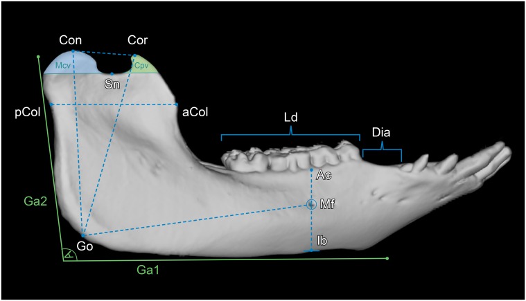
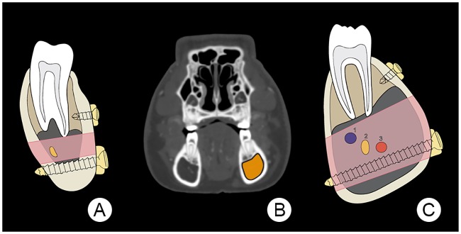
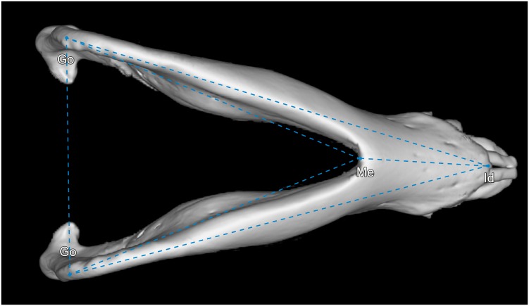
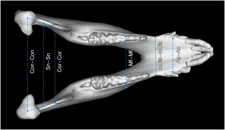
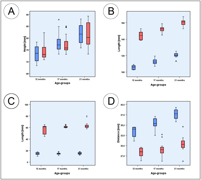
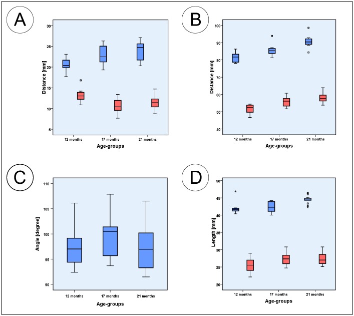
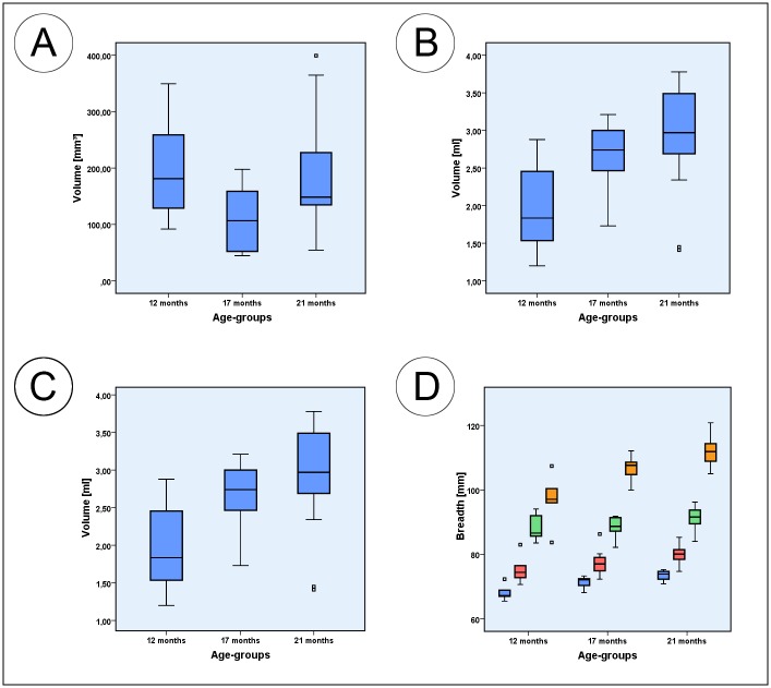
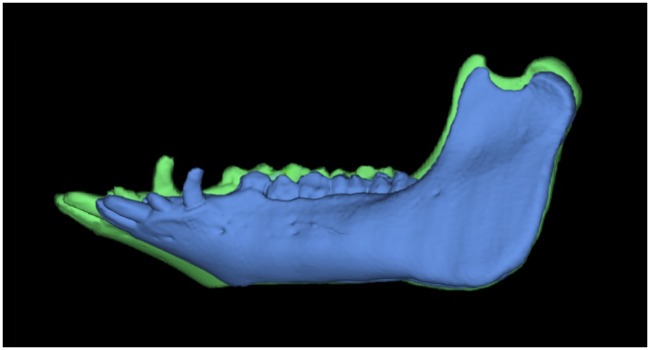
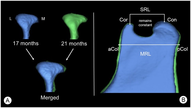
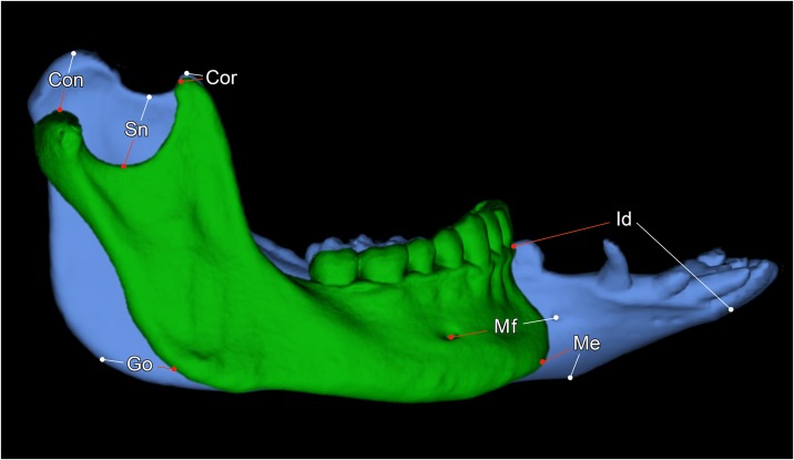
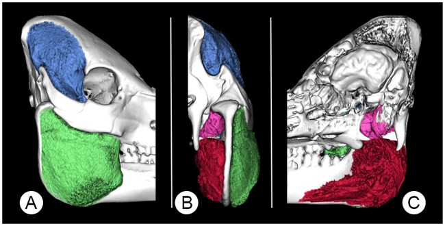
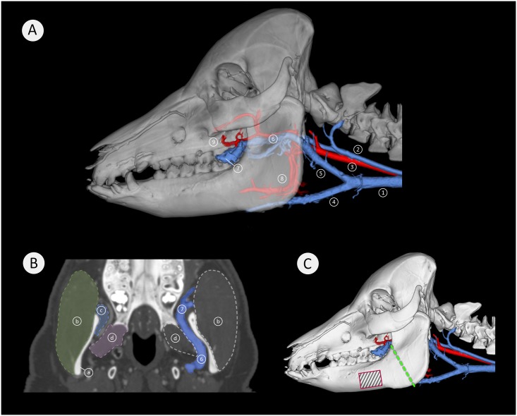
Over many decades, the Göttingen Minipig has been used as a large animal model in experimental surgical research of the mandible. Recently several authors have raised concerns over the use of the Göttingen Minipig in this research area, observing problems with post-operative wound healing and loosening implants. To reduce these complications during and after surgery and to improve animal welfare in mandibular surgery research, the present study elucidated how comparable the mandible of minipigs is to that of humans and whether these complications could be caused by specific anatomical characteristics of the minipigs' mandible, its masticatory muscles and associated vasculature. Twenty-two mandibular cephalometric parameters were measured on CT scans of Göttingen Minipigs aged between 12 and 21 months. Ultimately, we compared this data with human data reported in the scientific literature. In addition, image segmentation was used to determine the masticatory muscle morphology and the configuration of the mandibular blood vessels. Compared to data of humans, significant differences in the mandibular anatomy of minipigs were found. Of the 22 parameters measured only four were found to be highly comparable, whilst the others were not. The 3D examinations of the minipigs vasculature showed a very prominent deep facial vein directly medial to the mandibular ramus and potentially interfering with the sectional plane of mandibular distraction osteogenesis. Damage to this vessel could result in inaccessible bleeding. The findings of this study suggest that Göttingen Minipigs are not ideal animal models for experimental mandibular surgery research. Nevertheless if these minipigs are used the authors recommend that radiographic techniques, such as computed tomography, be used in the specific planning procedures for the mandibular surgical experiments. In addition, it is advisable to choose suitable age groups and customize implants based on the mandibular dimensions reported in this study.
几十年来,哥廷根小型猪一直被用作下颌骨实验外科研究的大型动物模型。最近,几位作者对在该研究领域使用哥廷根小型猪提出了担忧,观察到术后伤口愈合和植入物松动的问题。为了减少手术期间和手术后的这些并发症,并提高下颌骨手术研究中的动物福利,本研究阐明了小型猪的下颌骨与人类的下颌骨有多么相似,以及这些并发症是否可能是由小型猪下颌骨、其咀嚼肌及其相关血管的特定解剖结构特征引起的。对 12 至 21 月龄的哥廷根小型猪的 CT 扫描进行了 22 项下颌骨头颅测量参数测量。最终,我们将这些数据与科学文献中报告的人类数据进行了比较。此外,还使用图像分割来确定咀嚼肌形态和下颌血管结构。与人类数据相比,小型猪下颌骨解剖结构存在显著差异。在所测量的 22 个参数中,只有 4 个被认为高度相似,而其他参数则不然。小型猪血管的 3D 检查显示,在下颌支的内侧有一条非常突出的深部面静脉,可能会干扰下颌骨牵引成骨术的截面平面。如果损伤这条血管,可能会导致难以止血。本研究的结果表明,哥廷根小型猪不是实验性下颌骨手术研究的理想动物模型。尽管如此,如果使用这些小型猪,作者建议在具体的下颌骨手术实验规划程序中使用影像学技术,如计算机断层扫描。此外,建议根据本研究报告的下颌骨尺寸选择合适的年龄组,并定制植入物。