State Key Laboratory of Experimental HematologyInstitute of Hematology and Hospital of Blood Diseases, Chinese Academy of Medical Science and Peking Union Medical College, Tianjin, 300020, People's Republic of China.
Department of Pharmacy, National Clinical Research Center of Cancer, Key Laboratory of Cancer Prevention and Therapy, Tianjin's Clinical Research Center for Cancer, Tianjin Medical University Cancer Institute and Hospital, Tianjin, 300060, People's Republic of China.
J Hematol Oncol. 2019 Apr 25;12(1):46. doi: 10.1186/s13045-019-0723-8.
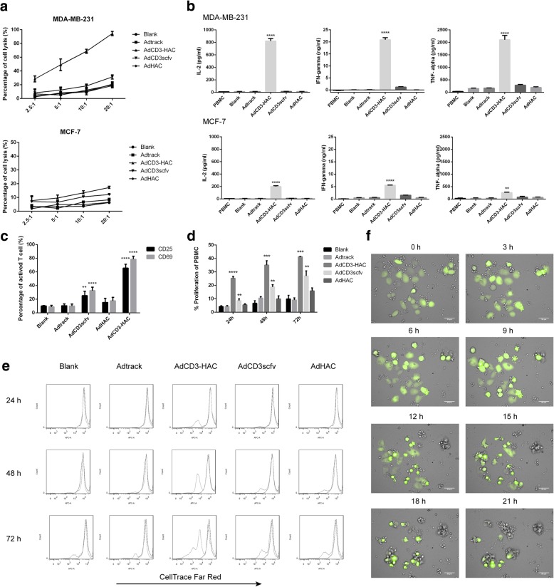
PD-1/PD-L1 blockade can confer durable benefits in the treatment of metastatic cancers, but the response rate remains modest and potential adverse effects occur sometimes. Concentrating immunotherapeutic agents at the site of disease was believed to break local immune tolerance and reduce systemic toxicity. E1A-engineered mesenchymal stromal cell (MSC.E1A) was an attractive transfer system that preferentially homing and treating cancer metastasis, through which the tumor cells were modified by locally replicated adenoviruses to release CD3-HAC, a bifunctional fusion protein that anti-CD3 scfv linked with high-affinity consensus (HAC) PD-1. Subsequently, CD3-HAC, wbich was bound on PD-L1-positive breast cancer cells, recruited T cells to exhibit a potent antitumor immunity incombination with immune checkpoint blockade.
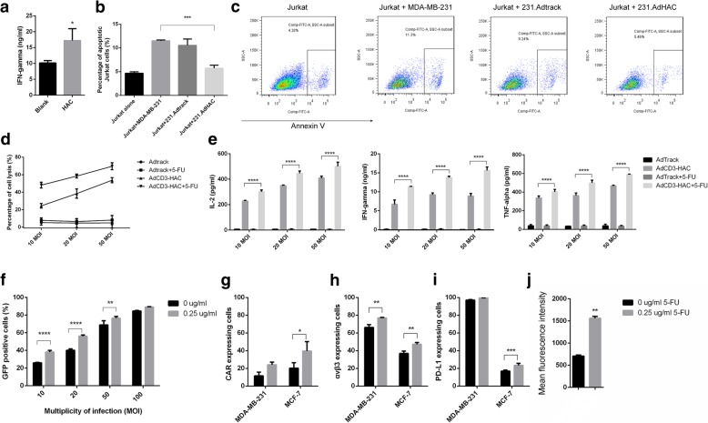
We constructed the CD3-HAC gene driven by human telomerase reverse transcriptase (hTERT) promoter into an adenoviral vector and the E1A gene into the lentiviral vector. The homing property of MSCs in vivo was analyzed with firefly luciferase-labeled MSCs (MSC.Luc) by bioluminescent imaging (BLI). The cytotoxicity of T cells induced by CD3-HAC towards PD-L1-positive cells was detected in vitro and in vivo in combination with 5-FU.
Our data suggest that CD3-HAC could specifically bind to PD-L1-positive tumor cells and induce lymphocyte-mediated lysis effectively both in vitro and in vivo. The intervention with HAC diminished the effects of PD-1/PD-L1 axis on T cells exposed to MDA-MB-231 cells and increased lymphocytes activation. MSCs infected by AdCD3-HAC followed by LentiR.E1A could specially migrate to metastasis of breast cancer and produce adenoviruses in the tumor sites. Furthermore, treatment with MSC.CD3-HAC.E1A in combination with 5-FU significantly inhibited the tumor growth in mice.
This adenovirus-loaded MSC.E1A system provides a promising strategy for the identification and elimination of metastasis with locally released immuno-modulator.
PD-1/PD-L1 阻断疗法可在转移性癌症的治疗中提供持久的疗效,但应答率仍然较低,并且有时会出现潜在的不良反应。将免疫治疗药物集中在疾病部位被认为可以打破局部免疫耐受并降低全身毒性。E1A 工程间充质基质细胞(MSC.E1A)是一种有吸引力的转移系统,它通过局部复制的腺病毒将肿瘤细胞修饰为释放 CD3-HAC,从而优先归巢和治疗癌症转移,CD3-HAC 是一种双功能融合蛋白,将抗 CD3 scfv 与高亲和力共识(HAC)PD-1 连接。随后,CD3-HAC 与 PD-L1 阳性乳腺癌细胞结合,招募 T 细胞在与免疫检查点阻断联合时表现出强大的抗肿瘤免疫。
我们构建了由人端粒酶逆转录酶(hTERT)启动子驱动的 CD3-HAC 基因到腺病毒载体和 E1A 基因到慢病毒载体。通过生物发光成像(BLI)分析 MSC 在体内的归巢特性。通过与 5-FU 联合,检测 CD3-HAC 诱导的 T 细胞对 PD-L1 阳性细胞的细胞毒性。
我们的数据表明,CD3-HAC 可以特异性结合 PD-L1 阳性肿瘤细胞,并在体外和体内有效诱导淋巴细胞介导的裂解。HAC 的干预减弱了 PD-1/PD-L1 轴对暴露于 MDA-MB-231 细胞的 T 细胞的影响,并增加了淋巴细胞的激活。感染 AdCD3-HAC 后再感染 LentiR.E1A 的 MSC 可以专门迁移到乳腺癌转移部位,并在肿瘤部位产生腺病毒。此外,与 5-FU 联合使用 MSC.CD3-HAC.E1A 治疗可显著抑制小鼠肿瘤生长。
这种负载腺病毒的 MSC.E1A 系统为识别和消除局部释放的免疫调节剂提供了一种有前途的策略。