WWAMI Medical Education Program, University of Washington School of Medicine, 1959 NE Pacific St, Seattle, WA, 98195, USA.
St Joseph Regional Medical Center, 415 6th St, Lewiston, ID, 83501, USA.
BMC Med Imaging. 2019 Apr 27;19(1):31. doi: 10.1186/s12880-019-0333-5.
A pseudoaneurysm occurs as the result of a contained rupture of an arterial wall, yielding a perfused sac that communicates with the arterial lumen. Pseudoaneurysm of an intercostal artery is an extremely rare event but it carries with it a significant risk of rupture and subsequent hemothorax. It must be considered as a potential complication of thoracentesis.
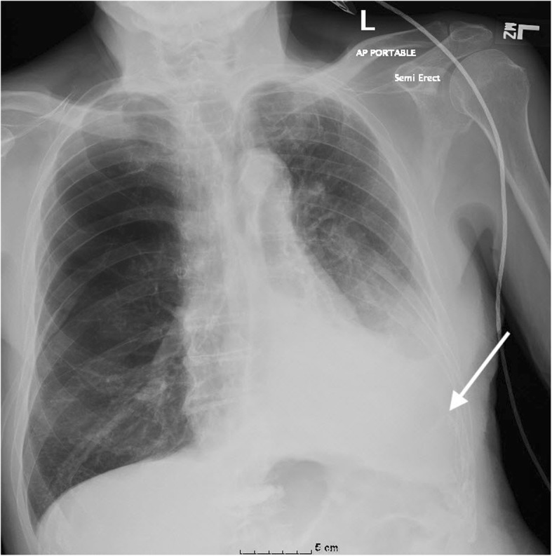
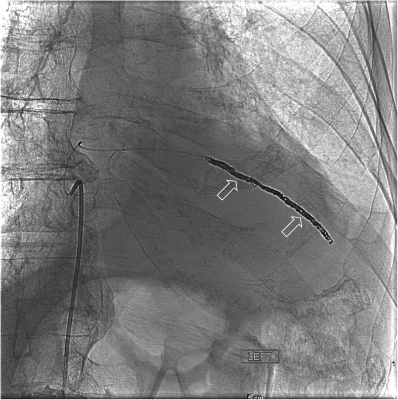
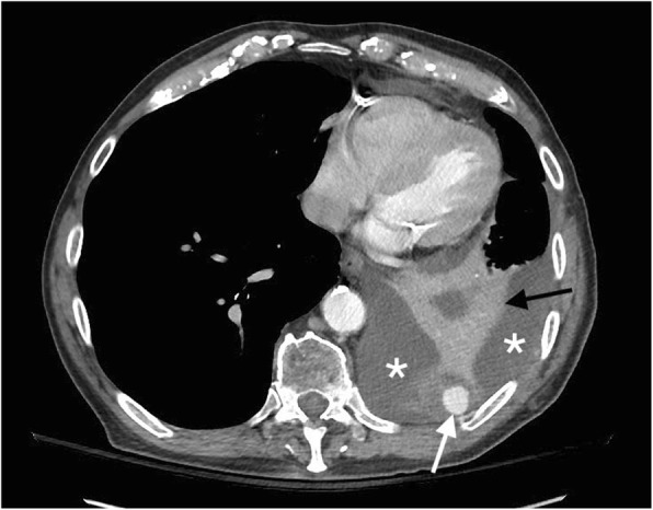
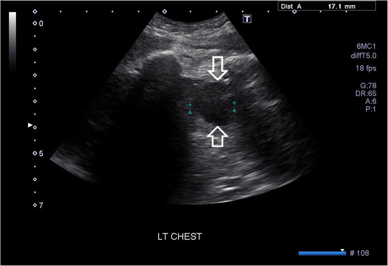
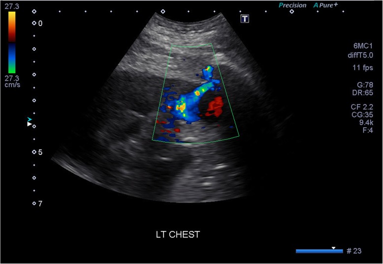
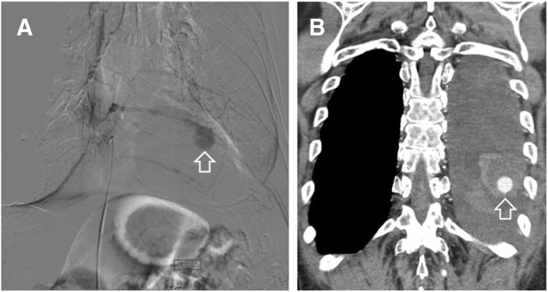
Here, we report a rare case of an intercostal artery pseudoaneurysm following thoracentesis in an 82-year old male. The patient presented with respiratory distress 1 day after a therapeutic thoracentesis had been performed. Computed tomography (CT) with contrast revealed a left intercostal pseudoaneurysm with hemothorax and adjacent compressive atelectasis. Doppler ultrasound revealed bidirectional blood flow in the pseudoaneurysm sac. An intercostal arteriogram and thoracic aortogram aided in confirmation of the pseudoaneurysm and successful treatment with coil embolization.
An intercostal pseudoaneurysm complication following thoracentesis is very rare but important to rule out as a possible cause of hemothorax after the procedure. Capturing this finding with the aid of multiple imaging modalities allowed for diagnostic certainty and rapid treatment with coil embolization, leading to a successful patient recovery.
假性动脉瘤是动脉壁破裂后被包裹而形成的,与动脉腔相通的一个充满血液的囊袋。肋间动脉假性动脉瘤极为罕见,但有破裂并随后导致血胸的重大风险。在进行胸腔穿刺时,必须将其视为一种潜在的并发症。
在此,我们报告一例 82 岁男性在胸腔穿刺后发生肋间动脉假性动脉瘤的罕见病例。患者在接受治疗性胸腔穿刺后 1 天出现呼吸窘迫。对比增强 CT 显示左侧肋间假性动脉瘤伴血胸和相邻压迫性肺不张。多普勒超声显示假性动脉瘤内双向血流。肋间动脉造影和胸主动脉造影有助于确认假性动脉瘤,并成功采用线圈栓塞治疗。
胸腔穿刺后发生肋间假性动脉瘤并发症非常罕见,但作为术后血胸的可能原因,需要排除。借助多种影像学手段捕捉到这一发现,有助于明确诊断,并迅速采用线圈栓塞治疗,使患者成功康复。