European Institute for Molecular Imaging (EIMI), University of Münster, Münster, Germany.
Imaging Neuroinflammation in Neurodegenerative Diseases (INMIND) EU FP7 consortium.
Theranostics. 2019 Feb 20;9(6):1523-1537. doi: 10.7150/thno.32461. eCollection 2019.
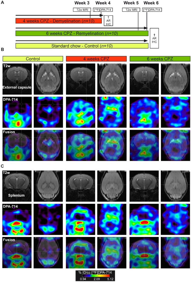
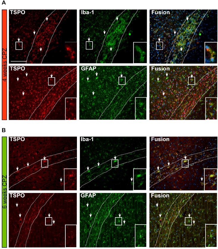
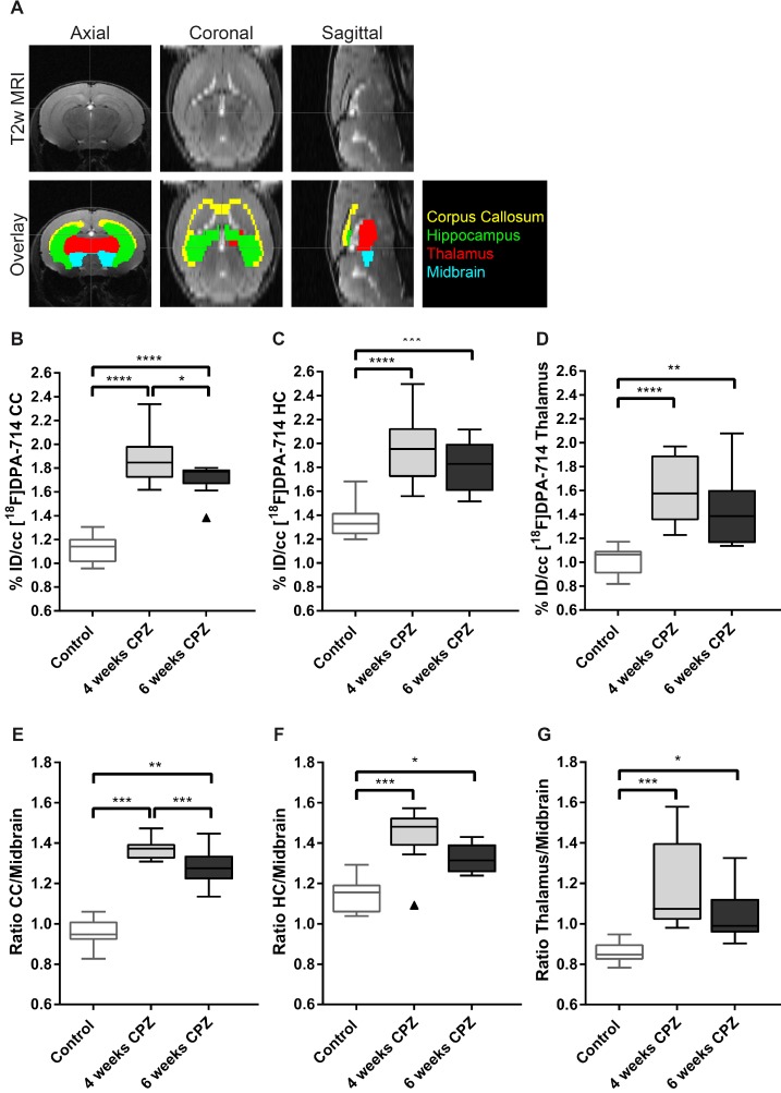
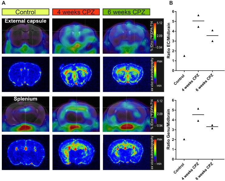
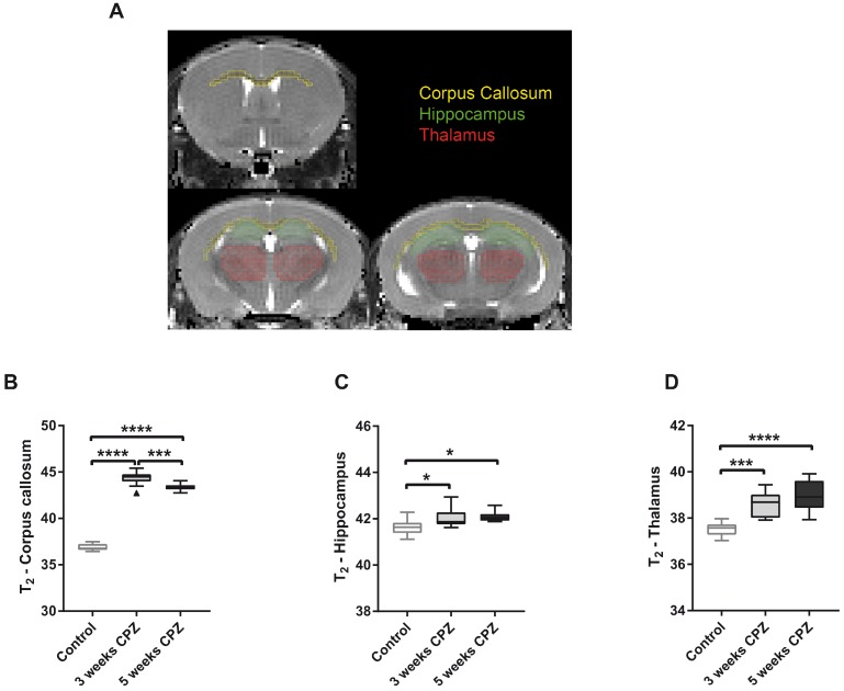
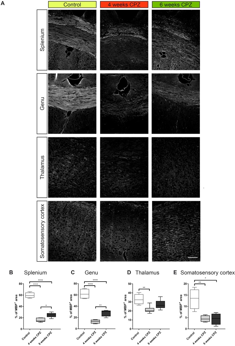
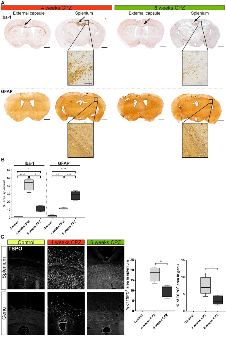
: Activation and dysregulation of innate, adaptive and resident immune cells in response to damage determine the pathophysiology of demyelinating disorders. Among the plethora of involved cells, microglia/macrophages and astrocytes play an important role in the pathogenesis of demyelinating disorders. The in-depth investigation of the spatio-temporal profile of these cell types may inform about the exact disease state and localization as well as may allow to monitor therapeutic modulation of the components of the neuroinflammatory response during the course of multiple sclerosis (MS). In this study, we aimed to non-invasively decipher the degree and temporal profile of neuroinflammation (TSPO - [F]DPA-714 PET) in relation to selected magnetic resonance imaging (MRI) parameters (T maps) in the cuprizone (CPZ)-induced model of demyelination. Methods: C57Bl6 () mice were fed with a standard chow mixed with 0.2% (w/w) CPZ for 4 (; demyelination) and 6 weeks (; spontaneous remyelination). The degree of neuroinflammation at de- and remyelination was assessed by [F]DPA-714 PET, multi-echo T MRI, autoradiography and immunohistochemistry. : CPZ-induced brain alterations were confirmed by increase of T relaxation times in both white and grey matter after 3 and 5 weeks of CPZ. Peak [F]DPA-714 was found in the corpus callosum (CC, white matter), the hippocampus (HC, grey matter) and thalamus (grey matter) after 4 weeks of CPZ treatment and declined after 6 weeks of CPZ. autoradiography and dedicated immunofluorescence showed demyelination/remyelination with corresponding increased/decreased TSPO levels in the CC and hippocampus, confirming the spatial distribution of [F]DPA-714 . The expression of TSPO microglia and astrocytes is time-dependent in this model. Microglia predominantly express TSPO at demyelination, while the majority of astrocytes express TSPO during remyelination. The combination of PET- and MRI-based imaging biomarkers demonstrated the regional and temporal development of the CPZ model-associated neuroinflammatory response in grey and white matter regions. : The combination of [F]DPA-714 PET and T mapping may allow to further elucidate the regional and temporal profile of inflammatory signals depending on the myelination status, although the underlying inflammatory microenvironment changes. A combination of the described imaging biomarkers may facilitate the development of patient-tailored strategies for immunomodulatory and neuro-restorative therapies in MS.
: 先天、适应性和固有免疫细胞的激活和失调对脱髓鞘疾病的发病机制产生反应。在众多涉及的细胞中,小胶质细胞/巨噬细胞和星形胶质细胞在脱髓鞘疾病的发病机制中起着重要作用。深入研究这些细胞类型的时空特征可能有助于了解确切的疾病状态和定位,并可能允许在多发性硬化症(MS)的病程中监测神经炎症反应成分的治疗调节。在这项研究中,我们旨在通过非侵入性方法破译神经炎症的程度和时间谱(TSPO-[F]DPA-714 PET)与杯状藻(CPZ)诱导的脱髓鞘模型中选定的磁共振成像(MRI)参数(T 图谱)之间的关系。方法:C57Bl6()小鼠用标准饲料喂养,饲料中混合 0.2%(w/w)CPZ 喂养 4 周(脱髓鞘)和 6 周(自发髓鞘再生)。通过[F]DPA-714 PET、多回波 T MRI、放射自显影和免疫组织化学评估脱髓鞘和髓鞘再生时的神经炎症程度。:CPZ 诱导的脑改变通过 CPZ 治疗 3 周和 5 周后白质和灰质 T 弛豫时间的增加得到证实。CPZ 治疗 4 周后,在胼胝体(CC,白质)、海马(HC,灰质)和丘脑(灰质)中发现[F]DPA-714 的峰值,并在 CPZ 治疗 6 周后下降。放射自显影和专门的免疫荧光显示 CC 和海马的脱髓鞘/髓鞘再生,相应地 TSPO 水平增加/减少,证实了[F]DPA-714 的空间分布。在该模型中,TSPO 小胶质细胞和星形胶质细胞的表达具有时间依赖性。小胶质细胞在脱髓鞘时主要表达 TSPO,而大多数星形胶质细胞在髓鞘再生时表达 TSPO。PET 和 MRI 为基础的成像生物标志物的组合显示了 CPZ 模型相关神经炎症反应在灰质和白质区域的区域和时间发展。:[F]DPA-714 PET 和 T 映射的组合可能允许进一步阐明炎症信号的区域和时间谱,这取决于髓鞘状态,尽管炎症微环境发生了变化。描述的成像生物标志物的组合可能有助于为多发性硬化症的免疫调节和神经修复治疗制定针对患者的策略。