Faculty of Health Sciences, Institute of Medical, Pharmaceutical and Health Sciences, Kanazawa University, Kanazawa, Japan.
Section of Radilogical Technology, Department of Medical Technology, Kanazawa Medical University Hospital, Uchinada, Kahoku, Japan.
J Appl Clin Med Phys. 2019 Jun;20(6):199-205. doi: 10.1002/acm2.12597. Epub 2019 May 2.
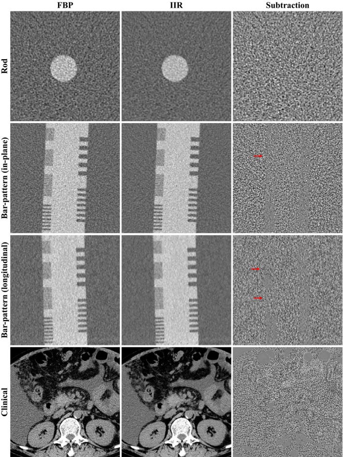
The purpose of this study is to evaluate the physical image quality of a commercially available image-based iterative reconstruction (IIR) system for two object contrasts to resemble a soft tissue (60 HU) and an enhanced vessel (270 HU), and compare the results with those of filtered back projection (FBP) and iterative reconstruction (IR). A 192-slice computed tomography (CT) scanner was used for data acquisitions. IIR images were processed from the FBP images. Task-based in-plane transfer function (TTF) and slice sensitivity profile (SSP ) were measured from rod objects inside of a 25-cm diameter water phantom at four dose levels (2.5, 5, 10, and 20 mGy). Noise power spectrum (NPS) was measured from the water-only part. System performance (SP) function was calculated as TTF /NPS over FBP, IR, and IIR for comparison. In addition, an image subtraction was performed using images of rod objects, a bar-pattern phantom, and a clinical abdomen case to observe the noise reduction performance of IIR. As a results, IIR mostly preserved TTF and SSP of FBP, whereas IR exhibited enhanced TTF at 10 and 20 mGy for 60 HU contrast and at all doses for 270 HU contrast. SP of IIR at 2.5, 5, 10 mGy (half doses) were similar to those of FBP at 5, 10, 20 mGy, respectively. IR exhibited enhanced SP at medium to high frequencies. The subtracted images showed weak remained edge signals in the bar-pattern and abdominal images. In conclusion, IIR uniformly improved the task-based image quality of FBP over the entire frequency range, whereas IR improved the characteristics over medium to high frequencies. The dose reduction potential of IIR estimated from SP is approximately 50%, when allowing the slight signal reductions.
本研究旨在评估一种商用基于图像的迭代重建(IIR)系统的物理图像质量,该系统针对两种目标对比度进行评估,分别模拟软组织(60 HU)和增强血管(270 HU)。将结果与滤波反投影(FBP)和迭代重建(IR)进行比较。使用一台 192 层 CT 扫描仪进行数据采集。从 FBP 图像中处理 IIR 图像。在四个剂量水平(2.5、5、10 和 20 mGy)下,从直径 25cm 的水模体内部的棒状物体测量基于任务的平面内传递函数(TTF)和切片灵敏度分布(SSP)。从仅水部分测量噪声功率谱(NPS)。计算 TTF/NPS 作为 FBP、IR 和 IIR 的系统性能(SP)函数进行比较。此外,通过使用棒状物体图像、条带图案体模和临床腹部病例进行图像相减,观察 IIR 的降噪性能。结果,IIR 主要保留了 FBP 的 TTF 和 SSP,而 IR 在 60 HU 对比度下的 10 和 20 mGy 以及所有剂量下的 270 HU 对比度下表现出增强的 TTF。在 2.5、5、10 mGy(半剂量)时,IIR 的 SP 与 FBP 在 5、10、20 mGy 时的 SP 相似。IR 在中高频显示出增强的 SP。相减图像显示条带图案和腹部图像中残留的边缘信号较弱。总之,IIR 在整个频率范围内均匀地提高了 FBP 的基于任务的图像质量,而 IR 则提高了中高频的特性。从 SP 估计的 IIR 的剂量减少潜力约为 50%,同时允许信号略有减少。