Institute of Neuroscience, Newcastle University, Newcastle upon Tyne, NE1 7RU, United Kingdom; Interdisciplinary Computing and Complex BioSystems (ICOS), School of Computing Science, Newcastle University, Newcastle upon Tyne, NE1 5TG, United Kingdom.
Institute of Cellular Medicine, Newcastle University, Newcastle upon Tyne, NE1 7RU, United Kingdom; Newcastle Magnetic Resonance Centre, Newcastle University, Campus for Ageing and Vitality, Newcastle upon Tyne, NE4 5PL, United Kingdom.
J Affect Disord. 2019 Jun 15;253:224-231. doi: 10.1016/j.jad.2019.04.075. Epub 2019 Apr 18.
Lithium treatment is associated with an increase in magnetic resonance imaging derived measures of white matter integrity, but the relationship between the spatial distribution of brain lithium and white matter integrity is unknown.
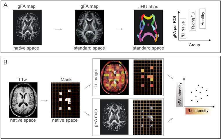
Euthymic patients with bipolar disorder receiving lithium (n = 12) and those on other medications but naïve to lithium (n = 17) underwent diffusion imaging alongside matched healthy controls (n = 16). Generalised fractional anisotropy (gFA) within white matter was compared between groups using a standard space white matter atlas. Lithium-treated patients underwent novel multinuclear lithium magnetic resonance imaging (Li-MRI) to determine the relative lithium concentration across the brain. The relationship between Li-MRI signal intensity and gFA was investigated at the resolution of the Li-MRI sequence in native space.
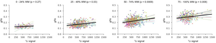
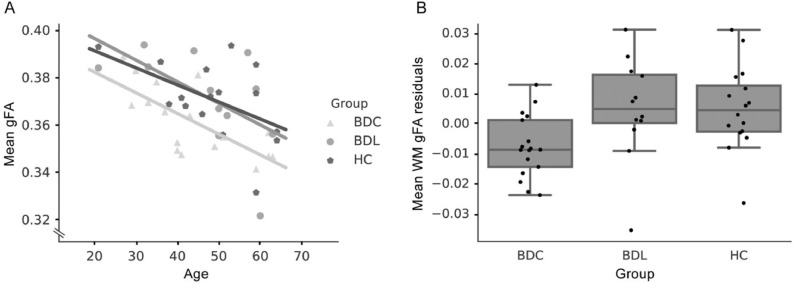
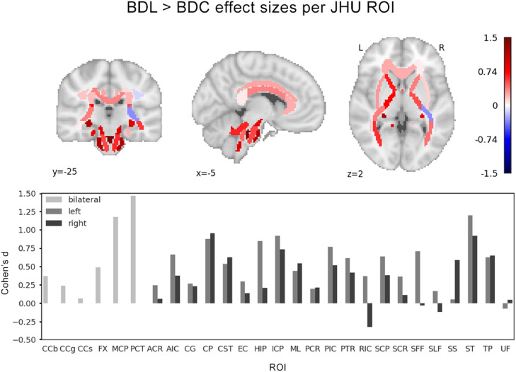
Lithium-treated bipolar disorder and healthy control groups had higher mean white matter gFA than the bipolar disorder group treated with other medications (t = 2.5, p < 0.05; t = 2.7, p < 0.03, respectively). No differences in gFA were found between patients taking lithium and healthy controls (t = 0.02, p = 1). These effects were seen consistently across most regions in the white matter atlas. In the lithium-treated group, a significant effect of the Li-MRI signal in predicting the gFA (p < 0.01) was identified in voxels containing over 50% white matter.
Cross-sectional evaluation of a relatively small cohort.
The higher gFA values observed in the lithium-treated bipolar disorder group suggests that long-term lithium is associated with greater white matter integrity. Our novel analysis supports this further, showing a positive association between white matter gFA and the spatial distribution of lithium.
锂治疗与磁共振成像(MRI)衍生的白质完整性测量值增加有关,但大脑内锂的空间分布与白质完整性之间的关系尚不清楚。
接受锂治疗的双相情感障碍稳定期患者(n=12)和接受其他药物治疗但尚未接受锂治疗的患者(n=17)与匹配的健康对照组(n=16)一起接受弥散成像。使用标准空间白质图谱比较各组白质内的广义各向异性分数(gFA)。对锂治疗患者进行新型多核锂磁共振成像(Li-MRI)以确定大脑内的相对锂浓度。在原生空间中以 Li-MRI 序列的分辨率,研究 Li-MRI 信号强度与 gFA 之间的关系。
锂治疗的双相情感障碍和健康对照组的平均白质 gFA 均高于接受其他药物治疗的双相情感障碍组(t=2.5,p<0.05;t=2.7,p<0.03)。锂治疗的患者与健康对照组之间 gFA 无差异(t=0.02,p=1)。这些效应在白质图谱的大多数区域中均一致存在。在锂治疗组中,Li-MRI 信号对 gFA 的预测存在显著影响(p<0.01),在包含超过 50%白质的体素中。
对相对较小队列的横断面评估。
锂治疗的双相情感障碍组观察到的 gFA 值较高,提示长期锂治疗与更大的白质完整性有关。我们的新分析进一步支持了这一点,显示白质 gFA 与锂的空间分布之间存在正相关。