Department of Radiology, Qilu Hospital of Shandong University, Jinan, Shandong, China (mainland).
Department of Radiology, Laigang Hospital Affiliated to Taishan Medical University, Laiwu, Shandong, China (mainland).
Med Sci Monit. 2019 May 5;25:3321-3328. doi: 10.12659/MSM.913439.
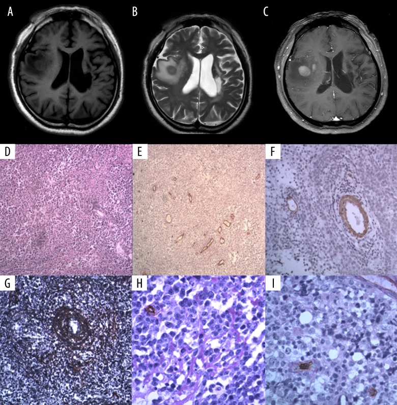
BACKGROUND This study aimed to compare the magnetic resonance imaging (MRI) findings of primary diffuse large B-cell lymphoma (DLBCL) of the central nervous system (CNS) with delayed contrast enhancement and histological microvessel density (MVD). T1-weighted and T2-weighted contrast-enhanced and non-enhanced brain imaging were used. CNS lymphoma tissue was evaluated using primary antibodies to endothelial cells and smooth muscle cells, and histochemical staining for reticulin fibers and basement membrane, which allowed quantification of the MVD. MATERIAL AND METHODS Twenty-one patients with histologically confirmed primary DLBCL of the CNS underwent pre-contrast-enhanced and postcontrast-enhanced MRI. Histology of the CNS lymphoma tissue included immunohistochemical staining with antibodies to CD34 for vascular endothelial cells and alpha smooth muscle actin (ASMA) for vascular smooth muscle cells, and histochemical staining included periodic acid-Schiff (PAS) and silver staining for reticulin fibers to evaluate microvessel density (MVD). RESULTS In primary DLBCL of the CNS, a positive correlation was found between the degree of necrosis and the size of the lymphoma (r=0.546, P=0.01). Delayed imaging enhancement was significantly correlated with the number of mature vessels, MVD, basement membrane, and reticulin fibers (r=0.593, 0.466, 0.446 and 0.497, respectively). Standardized ß regression coefficient analysis showed that the MVD, PAS-positive structures, the number of mature vessels, and reticulin fibers, were significantly associated with delayed enhancement on MRI (ß values, 0.425, 0.409, 0.295, and 0.188, respectively). CONCLUSIONS In primary DLBCL of the CNS, delayed imaging enhancement on MRI may be due to reduced neovascularization and vascular infiltration by lymphoma cells.
背景 本研究旨在比较原发性中枢神经系统(CNS)弥漫性大 B 细胞淋巴瘤(DLBCL)的磁共振成像(MRI)表现与延迟对比增强和组织微血管密度(MVD)。使用 T1 加权和 T2 加权对比增强和非增强脑成像。使用针对内皮细胞和平滑肌细胞的主要抗体以及网状纤维和基底膜的组织化学染色评估 CNS 淋巴瘤组织,从而可以定量评估 MVD。
材料与方法 21 例经组织学证实的原发性 CNS DLBCL 患者接受了增强前和增强后 MRI 检查。CNS 淋巴瘤组织的组织学包括针对血管内皮细胞的 CD34 抗体的免疫组织化学染色和针对血管平滑肌细胞的α平滑肌肌动蛋白(ASMA)的免疫组织化学染色,以及包括过碘酸希夫(PAS)和银染色的组织化学染色,以评估微血管密度(MVD)。
结果 在原发性 CNS DLBCL 中,坏死程度与淋巴瘤的大小呈正相关(r=0.546,P=0.01)。延迟成像增强与成熟血管的数量、MVD、基底膜和网状纤维显著相关(r=0.593、0.466、0.446 和 0.497)。标准化β回归系数分析显示,MVD、PAS 阳性结构、成熟血管的数量和网状纤维与 MRI 上的延迟增强显著相关(β值分别为 0.425、0.409、0.295 和 0.188)。
结论 在原发性 CNS DLBCL 中,MRI 上的延迟增强可能是由于新生血管减少和淋巴瘤细胞的血管浸润所致。