• 文献检索
  • 文档翻译
  • 深度研究
  • 学术资讯
  • Suppr Zotero 插件Zotero 插件
  • 邀请有礼
  • 套餐&价格
  • 历史记录
应用&插件
Suppr Zotero 插件Zotero 插件浏览器插件Mac 客户端Windows 客户端微信小程序
定价
高级版会员购买积分包购买API积分包
服务
文献检索文档翻译深度研究API 文档MCP 服务
关于我们
关于 Suppr公司介绍联系我们用户协议隐私条款
关注我们

Suppr 超能文献

核心技术专利:CN118964589B侵权必究
粤ICP备2023148730 号-1Suppr @ 2026

文献检索

告别复杂PubMed语法,用中文像聊天一样搜索,搜遍4000万医学文献。AI智能推荐,让科研检索更轻松。

立即免费搜索

文件翻译

保留排版,准确专业,支持PDF/Word/PPT等文件格式,支持 12+语言互译。

免费翻译文档

深度研究

AI帮你快速写综述,25分钟生成高质量综述,智能提取关键信息,辅助科研写作。

立即免费体验

磁共振成像评估小鼠胰腺导管腺癌中棕色脂肪组织与恶病质之间的关联

MRI Assessment of Associations between Brown Adipose Tissue and Cachexia in Murine Pancreatic Ductal Adenocarcinoma.

作者信息

Zhang Yaqi, Hu Su, Shangguan Junjie, Pan Liang, Zhou Xin, Yaghmai Vahid, Velichko Yuri, Hu Chunhong, Yang Jia, Zhang Zhuoli

机构信息

Department of Radiology, Feinberg School of Medicine, Northwestern University, Chicago, IL, USA.

Department of Radiology, The First Affiliated Hospital of Soochow University, Suzhou, Jiangsu, China.

出版信息

Intern Med Open Access. 2019;9(1). doi: 10.4172/2165-8048.1000301. Epub 2019 Feb 8.

DOI:10.4172/2165-8048.1000301
PMID:31073508
原文链接:https://pmc.ncbi.nlm.nih.gov/articles/PMC6502254/
Abstract

OBJECTIVE

As the major thermogenic tissue in body, the brown adipose tissue (BAT) was recently identified as an important factor to induce the rapid weight loss and malnutrition in malignancy. Current methods for detecting and quantifying brown adipose tissue (BAT) are in limited use. The aim of this study was to evaluate the changes of BAT tissue and its function in the development of pancreatic ductal adenocarcinoma (PDAC) by using magnetic resonance imaging (MRI).

METHODS

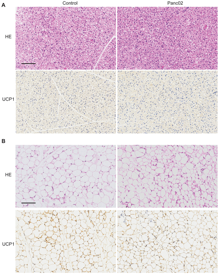
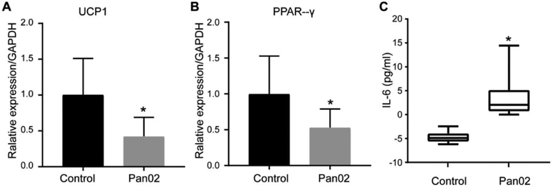
Ten-week-old female C57BL/6 mice were inoculated orthotopically with Pan02 tumor cells. R2* maps and two-point Dixon MRI were performed weekly for evaluation of BAT function and volume, respectively. The T2-weighted MRI was applied weekly for monitoring tumor growth. Meanwhile, the body weight was measured daily as another indication of malnutrition. The UCP1 levels in BAT and white adipose tissue (WAT) were assessed. The serum IL-6 was also measured as the biomarker of cancer-associated cachexia.

RESULTS

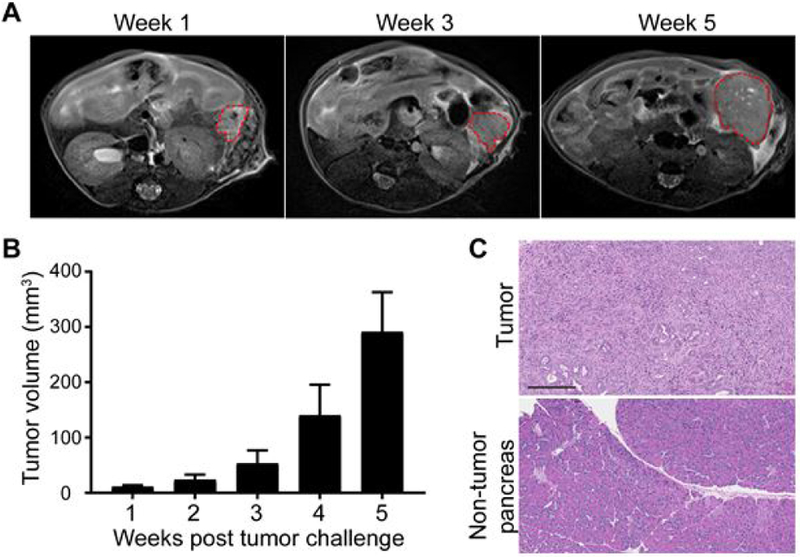
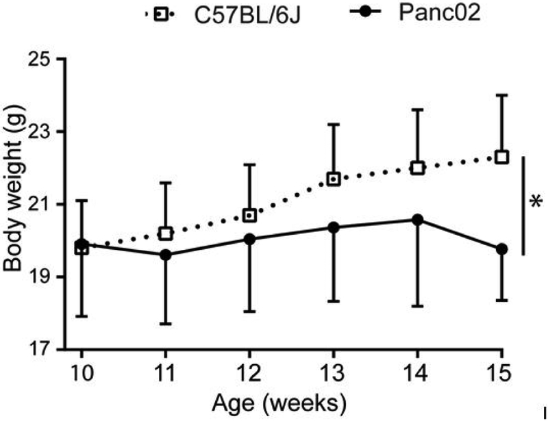
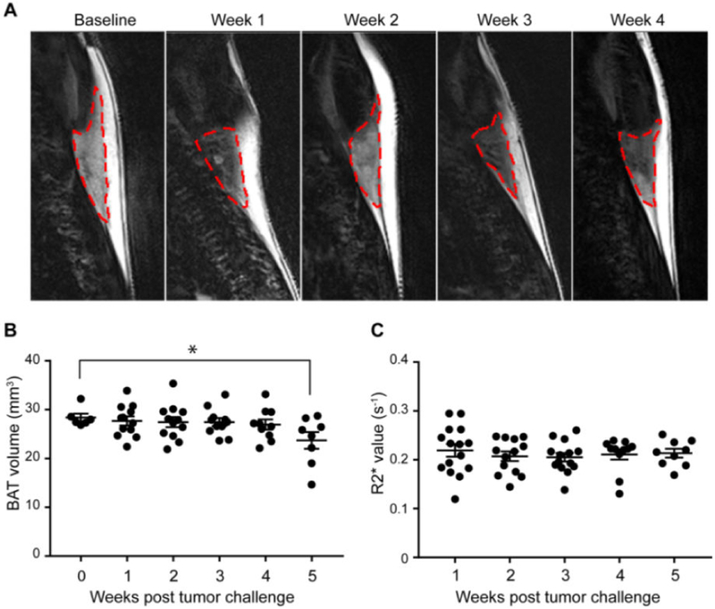
T2-weighted MRI indicated the rapid tumor growth from week 3 to week 5 after tumor cell inoculation. The water-fat separated MRI could clearly identify and quantify the BAT. The function and volume of BAT could be monitored by weekly MRI measurement in tumor-bearing mice. The total body weights of PDAC tumor-bearing mice were relatively stable, however, was significantly lower than that of control C57BL/6 mice.

CONCLUSION

The results of this study demonstrated the feasibility of detection and quantification of BAT by MRI during the development of pancreatic cancer.

摘要

目的

棕色脂肪组织(BAT)作为机体主要的产热组织,最近被确定为恶性肿瘤中导致快速体重减轻和营养不良的一个重要因素。目前检测和定量棕色脂肪组织(BAT)的方法应用有限。本研究的目的是利用磁共振成像(MRI)评估胰腺导管腺癌(PDAC)发生发展过程中BAT组织及其功能的变化。

方法

将Pan02肿瘤细胞原位接种于10周龄雌性C57BL/6小鼠。每周分别进行R2*图和两点 Dixon MRI检查,以评估BAT功能和体积。每周应用T2加权MRI监测肿瘤生长。同时,每天测量体重作为营养不良的另一指标。评估BAT和白色脂肪组织(WAT)中解偶联蛋白1(UCP1)的水平。还检测血清白细胞介素-6(IL-6)作为癌症相关性恶病质的生物标志物。

结果

T2加权MRI显示肿瘤细胞接种后第3周~第5周肿瘤快速生长。水脂分离MRI能够清晰地识别和定量BAT。通过对荷瘤小鼠每周进行MRI测量可监测BAT的功能和体积。PDAC荷瘤小鼠的总体重相对稳定,然而,显著低于对照C57BL/6小鼠。

结论

本研究结果证明了在胰腺癌发生发展过程中利用MRI检测和定量BAT的可行性。

https://cdn.ncbi.nlm.nih.gov/pmc/blobs/6b76/6502254/095a3eee02b1/nihms-1017409-f0005.jpg
https://cdn.ncbi.nlm.nih.gov/pmc/blobs/6b76/6502254/e8762570a1ec/nihms-1017409-f0001.jpg
https://cdn.ncbi.nlm.nih.gov/pmc/blobs/6b76/6502254/754ae8cad4dc/nihms-1017409-f0002.jpg
https://cdn.ncbi.nlm.nih.gov/pmc/blobs/6b76/6502254/b81040dab838/nihms-1017409-f0003.jpg
https://cdn.ncbi.nlm.nih.gov/pmc/blobs/6b76/6502254/3a580f6298da/nihms-1017409-f0004.jpg
https://cdn.ncbi.nlm.nih.gov/pmc/blobs/6b76/6502254/095a3eee02b1/nihms-1017409-f0005.jpg

相似文献

1
MRI Assessment of Associations between Brown Adipose Tissue and Cachexia in Murine Pancreatic Ductal Adenocarcinoma.磁共振成像评估小鼠胰腺导管腺癌中棕色脂肪组织与恶病质之间的关联
Intern Med Open Access. 2019;9(1). doi: 10.4172/2165-8048.1000301. Epub 2019 Feb 8.
2
Quantification of brown adipose tissue using synthetic magnetic resonance imaging: an experimental study with mice model.使用合成磁共振成像对棕色脂肪组织进行定量分析:一项小鼠模型实验研究。
Quant Imaging Med Surg. 2022 Jan;12(1):526-538. doi: 10.21037/qims-20-1344.
3
Quantification of brown and white adipose tissue based on Gaussian mixture model using water-fat and T2* MRI in adolescents.基于水脂和 T2*MRI 的高斯混合模型对青少年棕色和白色脂肪组织的定量研究。
J Magn Reson Imaging. 2017 Sep;46(3):758-768. doi: 10.1002/jmri.25632. Epub 2017 Jan 16.
4
MRI characterization of brown adipose tissue in obese and normal-weight children.肥胖和正常体重儿童棕色脂肪组织的MRI特征
Pediatr Radiol. 2015 Oct;45(11):1682-9. doi: 10.1007/s00247-015-3391-z. Epub 2015 Jun 20.
5
A method for the automatic segmentation of brown adipose tissue.一种用于棕色脂肪组织自动分割的方法。
MAGMA. 2016 Apr;29(2):287-99. doi: 10.1007/s10334-015-0517-0. Epub 2016 Jan 11.
6
Discrimination Between Brown and White Adipose Tissue Using a 2-Point Dixon Water-Fat Separation Method in Simultaneous PET/MRI.在同步PET/MRI中使用两点Dixon水脂分离法鉴别棕色脂肪组织和白色脂肪组织
J Nucl Med. 2015 Nov;56(11):1742-7. doi: 10.2967/jnumed.115.160770. Epub 2015 Aug 13.
7
MRI characterization of brown adipose tissue under thermal challenges in normal weight, overweight, and obese young men.正常体重、超重和肥胖年轻男性在热挑战下的棕色脂肪组织的 MRI 特征。
J Magn Reson Imaging. 2018 Apr;47(4):936-947. doi: 10.1002/jmri.25836. Epub 2017 Aug 11.
8
MR signal-fat-fraction analysis and T2* weighted imaging measure BAT reliably on humans without cold exposure.MR 信号脂肪分数分析和 T2* 加权成像测量 BAT 在不进行冷暴露的情况下可靠地应用于人体。
Metabolism. 2017 May;70:23-30. doi: 10.1016/j.metabol.2017.02.001. Epub 2017 Feb 8.
9
Observed changes in brown, white, hepatic and pancreatic fat after bariatric surgery: Evaluation with MRI.减重手术后棕色、白色、肝性和胰腺脂肪的观察性变化:MRI 评估。
Eur Radiol. 2019 Feb;29(2):849-856. doi: 10.1007/s00330-018-5611-z. Epub 2018 Jul 30.
10
Measurement of brown adipose tissue mass using a novel dual-echo magnetic resonance imaging approach: a validation study.采用新型双回波磁共振成像方法测量棕色脂肪组织质量:一项验证研究。
Metabolism. 2013 Aug;62(8):1189-98. doi: 10.1016/j.metabol.2013.03.002. Epub 2013 Apr 13.

引用本文的文献

1
Quantitative and Qualitative Radiological Assessment of Sarcopenia and Cachexia in Cancer Patients: A Systematic Review.癌症患者肌肉减少症和恶病质的定量与定性放射学评估:一项系统综述
J Pers Med. 2024 Feb 24;14(3):243. doi: 10.3390/jpm14030243.
2
Adipose Tissue Wasting as a Determinant of Pancreatic Cancer-Related Cachexia.脂肪组织消耗作为胰腺癌相关恶病质的一个决定因素。
Cancers (Basel). 2022 Sep 29;14(19):4754. doi: 10.3390/cancers14194754.
3
The evolving view of thermogenic fat and its implications in cancer and metabolic diseases.

本文引用的文献

1
Altered exocrine function can drive adipose wasting in early pancreatic cancer.外分泌功能改变可导致早期胰腺癌的脂肪消耗。
Nature. 2018 Jun;558(7711):600-604. doi: 10.1038/s41586-018-0235-7. Epub 2018 Jun 20.
2
Peroxisome Proliferator Activated Receptor Gamma Controls Mature Brown Adipocyte Inducibility through Glycerol Kinase.过氧化物酶体增殖物激活受体γ通过甘油激酶控制成熟棕色脂肪细胞的诱导能力。
Cell Rep. 2018 Jan 16;22(3):760-773. doi: 10.1016/j.celrep.2017.12.067.
3
Cancer-associated cachexia.癌症相关性恶病质。
生热脂肪的不断演变的观点及其在癌症和代谢性疾病中的意义。
Signal Transduct Target Ther. 2022 Sep 16;7(1):324. doi: 10.1038/s41392-022-01178-6.
4
Clinical Application of Bioextracts in Supporting the Reproductive System of Animals and Humans: Potential and Limitations.生物提取物在支持动物和人类生殖系统中的临床应用:潜力与局限
Evid Based Complement Alternat Med. 2022 Mar 27;2022:4766409. doi: 10.1155/2022/4766409. eCollection 2022.
5
Imaging modalities for diagnosis and monitoring of cancer cachexia.用于癌症恶病质诊断和监测的成像方式。
EJNMMI Res. 2021 Sep 23;11(1):94. doi: 10.1186/s13550-021-00834-2.
6
Role of Antioxidant Natural Products in Management of Infertility: A Review of Their Medicinal Potential.抗氧化天然产物在不孕症治疗中的作用:对其药用潜力的综述
Antioxidants (Basel). 2020 Oct 7;9(10):957. doi: 10.3390/antiox9100957.
Nat Rev Dis Primers. 2018 Jan 18;4:17105. doi: 10.1038/nrdp.2017.105.
4
Predictors of Pancreatic Cancer-Associated Weight Loss and Nutritional Interventions.胰腺癌相关体重减轻的预测因素及营养干预措施。
Pancreas. 2017 Oct;46(9):1152-1157. doi: 10.1097/MPA.0000000000000898.
5
Establishment and characterization of a novel murine model of pancreatic cancer cachexia.建立并鉴定一种新型胰腺癌恶病质小鼠模型。
J Cachexia Sarcopenia Muscle. 2017 Oct;8(5):824-838. doi: 10.1002/jcsm.12225. Epub 2017 Jul 20.
6
MR signal-fat-fraction analysis and T2* weighted imaging measure BAT reliably on humans without cold exposure.MR 信号脂肪分数分析和 T2* 加权成像测量 BAT 在不进行冷暴露的情况下可靠地应用于人体。
Metabolism. 2017 May;70:23-30. doi: 10.1016/j.metabol.2017.02.001. Epub 2017 Feb 8.
7
Mechanisms of metabolic dysfunction in cancer-associated cachexia.癌症恶病质相关代谢功能障碍的机制。
Genes Dev. 2016 Mar 1;30(5):489-501. doi: 10.1101/gad.276733.115.
8
TGF-β Blockade Reduces Mortality and Metabolic Changes in a Validated Murine Model of Pancreatic Cancer Cachexia.在经过验证的胰腺癌恶病质小鼠模型中,转化生长因子-β(TGF-β)阻断可降低死亡率并改善代谢变化。
PLoS One. 2015 Jul 14;10(7):e0132786. doi: 10.1371/journal.pone.0132786. eCollection 2015.
9
Elevation of circulating branched-chain amino acids is an early event in human pancreatic adenocarcinoma development.循环中支链氨基酸水平升高是人类胰腺腺癌发生过程中的早期事件。
Nat Med. 2014 Oct;20(10):1193-1198. doi: 10.1038/nm.3686. Epub 2014 Sep 28.
10
A switch from white to brown fat increases energy expenditure in cancer-associated cachexia.由白色脂肪向棕色脂肪的转变可增加癌症恶病质相关的能量消耗。
Cell Metab. 2014 Sep 2;20(3):433-47. doi: 10.1016/j.cmet.2014.06.011. Epub 2014 Jul 17.