• 文献检索
  • 文档翻译
  • 深度研究
  • 学术资讯
  • Suppr Zotero 插件Zotero 插件
  • 邀请有礼
  • 套餐&价格
  • 历史记录
应用&插件
Suppr Zotero 插件Zotero 插件浏览器插件Mac 客户端Windows 客户端微信小程序
定价
高级版会员购买积分包购买API积分包
服务
文献检索文档翻译深度研究API 文档MCP 服务
关于我们
关于 Suppr公司介绍联系我们用户协议隐私条款
关注我们

Suppr 超能文献

核心技术专利:CN118964589B侵权必究
粤ICP备2023148730 号-1Suppr @ 2026

文献检索

告别复杂PubMed语法,用中文像聊天一样搜索,搜遍4000万医学文献。AI智能推荐,让科研检索更轻松。

立即免费搜索

文件翻译

保留排版,准确专业,支持PDF/Word/PPT等文件格式,支持 12+语言互译。

免费翻译文档

深度研究

AI帮你快速写综述,25分钟生成高质量综述,智能提取关键信息,辅助科研写作。

立即免费体验

正常体重、超重和肥胖年轻男性在热挑战下的棕色脂肪组织的 MRI 特征。

MRI characterization of brown adipose tissue under thermal challenges in normal weight, overweight, and obese young men.

机构信息

Department of Medical Imaging, Ann & Robert H. Lurie Children's Hospital of Chicago, Chicago, Illinois, USA.

Department of Radiology, Feinberg School of Medicine, Northwestern University, Chicago, Illinois, USA.

出版信息

J Magn Reson Imaging. 2018 Apr;47(4):936-947. doi: 10.1002/jmri.25836. Epub 2017 Aug 11.

DOI:10.1002/jmri.25836
PMID:28801960
原文链接:https://pmc.ncbi.nlm.nih.gov/articles/PMC5808911/
Abstract

PURPOSE

To implement quantitative Dixon magnetic resonance imaging (MRI) methods for brown adipose tissue (BAT) characterization at inactive and cold-activated states in normal weight, overweight, and obese subjects. The hypotheses are that MRI characteristics of BAT would differentiate between nonobese and obese subjects, and activation of BAT in response to thermal challenges that are detected by MRI would be correlated with BAT activity measured by positron emission tomography / computed tomography (PET/CT).

MATERIALS AND METHODS

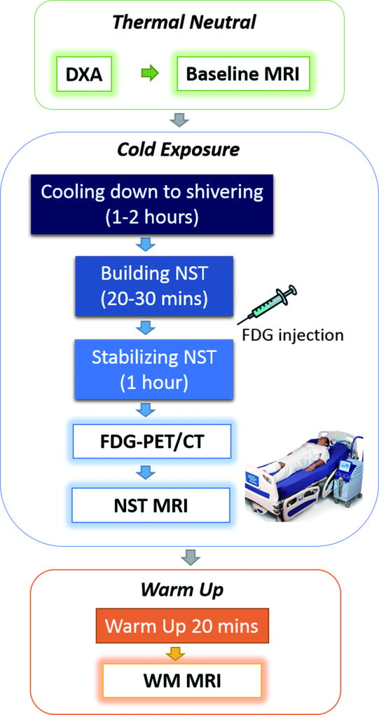
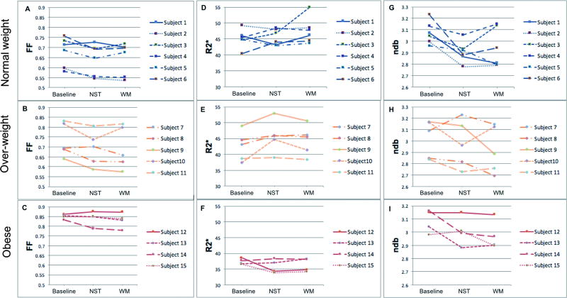
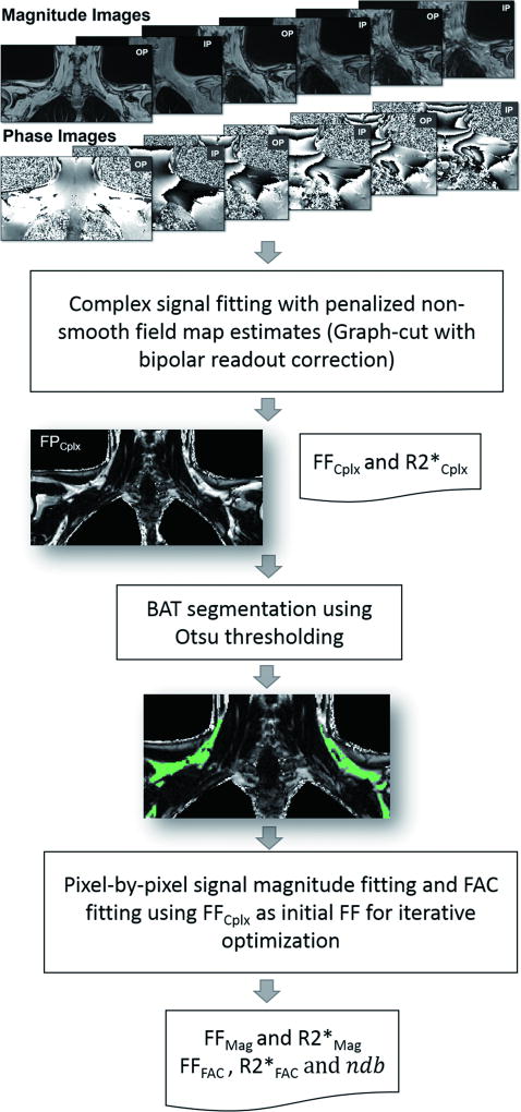
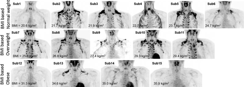
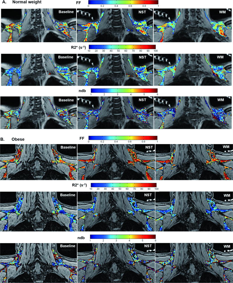
Fifteen male subjects (20.7 ± 1.5 years old) including six normal weight, five overweight, and four obese subjects participated in the study. A multiecho Dixon MRI sequence was performed on a 1.5T scanner. MRI was acquired under thermoneutral, nonshivering thermogenesis, and subsequent warm-up conditions. Fat fraction (FF), R2*, and the number of double bonds (ndb) were measured by solving an optimization problem that fits in- and out-of-phase MR signal intensities to the fat-water interference models. Imaging acquisition and postprocessing were performed by two MRI physicists. In each subject, Dixon MRI measurements of FF, R2*, and ndb were calculated for each voxel within all BAT regions of interest (ROIs) under each thermal condition. Mean FF, R2*, and ndb were compared between nonobese (ie, normal-weight/overweight) and obese subjects using the two-sample t-test. Receiver operating characteristic (ROC) analyses were performed to differentiate nonobese vs. obese subjects. BAT MRI measurement changes in response to thermal condition changes were compared with hypermetabolic BAT volume/activity measured by PET/CT using the Pearson's correlation. In addition, BAT MRI measurements were compared with body adiposity using the Pearson's correlation. P < 0.05 was considered statistically significant.

RESULTS

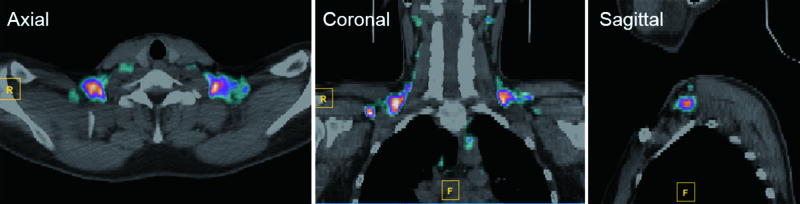
Obese subjects showed higher FF and lower R2* than nonobese subjects under all three thermal conditions (P < 0.01). ROC analyses demonstrated that FF and R2* were excellent predictors for the differentiation of nonobese from obese subjects (100% specificity and 100% sensitivity). FF changes under thermal challenges were correlated with hypermetabolic BAT volume (r = -0.55, P = 0.04 during activation, and r = 0.72, P = 0.003 during deactivation), and with BAT activity (r = 0.69, P = 0.006 during deactivation), as measured by PET/CT. FF and R2* under all three thermal conditions were highly correlated with body adiposity (P ≤ 0.002).

CONCLUSION

MRI characteristics of BAT differentiated between nonobese and obese subjects in both inactivated and activated states. BAT activation detected by Dixon MRI in response to thermal challenges were correlated with glucose uptake of metabolically active BAT.

LEVEL OF EVIDENCE

1 Technical Efficacy: Stage 3 J. Magn. Reson. Imaging 2018;47:936-947.

摘要

目的

在正常体重、超重和肥胖受试者的静息和冷激活状态下,实现用于棕色脂肪组织 (BAT) 特征描述的定量 Dixon 磁共振成像 (MRI) 方法。假设 MRI 特征可以区分非肥胖和肥胖受试者,并且通过 MRI 检测到的对热挑战的 BAT 激活将与通过正电子发射断层扫描/计算机断层扫描 (PET/CT) 测量的 BAT 活性相关。

材料与方法

15 名男性受试者(20.7±1.5 岁),包括 6 名正常体重者、5 名超重者和 4 名肥胖者,参加了这项研究。在 1.5T 扫描仪上进行多回波 Dixon MRI 序列。MRI 在体温中性、非颤抖产热和随后的热身条件下采集。通过解决拟合同相位和反相位 MR 信号强度与脂肪-水干扰模型的优化问题,测量脂肪分数 (FF)、R2和双键数 (ndb)。每个受试者的所有 BAT 感兴趣区域 (ROI) 内的每个体素的 Dixon MRI 测量值均在每个热条件下进行计算。使用两样本 t 检验比较非肥胖(即正常体重/超重)和肥胖受试者之间的 FF、R2和 ndb 的平均值。使用接收器工作特性 (ROC) 分析来区分非肥胖与肥胖受试者。使用 Pearson 相关比较 BAT MRI 测量值在热条件变化下的变化与 PET/CT 测量的高代谢 BAT 体积/活性。此外,使用 Pearson 相关比较 BAT MRI 测量值与身体脂肪含量。P<0.05 被认为具有统计学意义。

结果

在所有三种热条件下,肥胖受试者的 FF 均高于非肥胖受试者,而 R2则低于非肥胖受试者(P<0.01)。ROC 分析表明,FF 和 R2是区分非肥胖和肥胖受试者的优秀预测指标(100%特异性和 100%敏感性)。在热挑战下,FF 变化与高代谢 BAT 体积(激活时 r=−0.55,P=0.04,失活时 r=0.72,P=0.003)和 BAT 活性(失活时 r=0.69,P=0.006)相关,通过 PET/CT 测量。在所有三种热条件下,FF 和 R2*与身体脂肪含量高度相关(P≤0.002)。

结论

在静息和激活状态下,BAT 的 MRI 特征可以区分非肥胖和肥胖受试者。对热挑战的 Dixon MRI 检测到的 BAT 激活与代谢活跃的 BAT 的葡萄糖摄取相关。

证据水平

1 技术功效:第 3 阶段 J. Magn. Reson. Imaging 2018;47:936-947.

https://cdn.ncbi.nlm.nih.gov/pmc/blobs/d6e7/5808911/923be9b8ae28/nihms928702f6.jpg
https://cdn.ncbi.nlm.nih.gov/pmc/blobs/d6e7/5808911/6f09fd46522f/nihms928702f1.jpg
https://cdn.ncbi.nlm.nih.gov/pmc/blobs/d6e7/5808911/a93f8768e770/nihms928702f2.jpg
https://cdn.ncbi.nlm.nih.gov/pmc/blobs/d6e7/5808911/c70d90fb8f31/nihms928702f3.jpg
https://cdn.ncbi.nlm.nih.gov/pmc/blobs/d6e7/5808911/c26066b18924/nihms928702f4.jpg
https://cdn.ncbi.nlm.nih.gov/pmc/blobs/d6e7/5808911/6eab0b138453/nihms928702f5.jpg
https://cdn.ncbi.nlm.nih.gov/pmc/blobs/d6e7/5808911/923be9b8ae28/nihms928702f6.jpg

相似文献

1
MRI characterization of brown adipose tissue under thermal challenges in normal weight, overweight, and obese young men.正常体重、超重和肥胖年轻男性在热挑战下的棕色脂肪组织的 MRI 特征。
J Magn Reson Imaging. 2018 Apr;47(4):936-947. doi: 10.1002/jmri.25836. Epub 2017 Aug 11.
2
MRI characteristics of supraclavicular brown adipose tissue in relation to cold-induced thermogenesis in healthy human adults.锁骨上棕色脂肪组织的 MRI 特征与健康成年人冷诱导产热的关系。
J Magn Reson Imaging. 2019 Oct;50(4):1160-1168. doi: 10.1002/jmri.26733. Epub 2019 Apr 4.
3
Identification of an optimal threshold for detecting human brown adipose tissue using receiver operating characteristic analysis of IDEAL MRI fat fraction maps.使用IDEAL MRI脂肪分数图的受试者工作特征分析来确定检测人体棕色脂肪组织的最佳阈值。
Magn Reson Imaging. 2018 Sep;51:61-68. doi: 10.1016/j.mri.2018.04.013. Epub 2018 Apr 26.
4
A pilot study on the correlation between fat fraction values and glucose uptake values in supraclavicular fat by simultaneous PET/MRI.锁骨上脂肪的 PET/MRI 同步检查中脂肪分数值与葡萄糖摄取值的相关性初步研究
Magn Reson Med. 2017 Nov;78(5):1922-1932. doi: 10.1002/mrm.26589. Epub 2017 Jan 23.
5
Characterizing active and inactive brown adipose tissue in adult humans using PET-CT and MR imaging.使用正电子发射断层扫描-计算机断层扫描(PET-CT)和磁共振成像(MR)对成年人体内的活性和非活性棕色脂肪组织进行表征。
Am J Physiol Endocrinol Metab. 2016 Jul 1;311(1):E95-E104. doi: 10.1152/ajpendo.00482.2015. Epub 2016 May 10.
6
Quantification of brown and white adipose tissue based on Gaussian mixture model using water-fat and T2* MRI in adolescents.基于水脂和 T2*MRI 的高斯混合模型对青少年棕色和白色脂肪组织的定量研究。
J Magn Reson Imaging. 2017 Sep;46(3):758-768. doi: 10.1002/jmri.25632. Epub 2017 Jan 16.
7
Discrimination Between Brown and White Adipose Tissue Using a 2-Point Dixon Water-Fat Separation Method in Simultaneous PET/MRI.在同步PET/MRI中使用两点Dixon水脂分离法鉴别棕色脂肪组织和白色脂肪组织
J Nucl Med. 2015 Nov;56(11):1742-7. doi: 10.2967/jnumed.115.160770. Epub 2015 Aug 13.
8
Imaging cold-activated brown adipose tissue using dynamic T2*-weighted magnetic resonance imaging and 2-deoxy-2-[18F]fluoro-D-glucose positron emission tomography.利用动态 T2*-加权磁共振成像和 2-脱氧-2-[18F]氟-D-葡萄糖正电子发射断层扫描成像技术对冷激活棕色脂肪组织进行成像。
Invest Radiol. 2013 Oct;48(10):708-14. doi: 10.1097/RLI.0b013e31829363b8.
9
Hybrid PET/MRI as a tool to detect brown adipose tissue: Proof of principle.混合正电子发射断层显像/磁共振成像作为检测棕色脂肪组织的工具:原理验证
Obes Res Clin Pract. 2015 Nov-Dec;9(6):613-7. doi: 10.1016/j.orcp.2015.05.004. Epub 2015 Jun 4.
10
MRI characterization of brown adipose tissue in obese and normal-weight children.肥胖和正常体重儿童棕色脂肪组织的MRI特征
Pediatr Radiol. 2015 Oct;45(11):1682-9. doi: 10.1007/s00247-015-3391-z. Epub 2015 Jun 20.

引用本文的文献

1
Cold exposure and thermoneutrality similarly reduce supraclavicular brown adipose tissue fat fraction in fasted young lean adults.寒冷暴露和热中性同样会降低禁食的年轻瘦成年人锁骨上棕色脂肪组织的脂肪含量。
FASEB J. 2025 Jan 15;39(1):e70307. doi: 10.1096/fj.202402415R.
2
The characterization of metabolic changes in adipose tissues and muscles due to different exercise intensities by Dixon in healthy young men.迪克森研究健康年轻男性不同运动强度下脂肪组织和肌肉代谢变化的特征。
Eur J Radiol. 2024 Aug;177:111559. doi: 10.1016/j.ejrad.2024.111559. Epub 2024 Jun 8.
3
Nipsnap1-A regulatory factor required for long-term maintenance of non-shivering thermogenesis.

本文引用的文献

1
MR signal-fat-fraction analysis and T2* weighted imaging measure BAT reliably on humans without cold exposure.MR 信号脂肪分数分析和 T2* 加权成像测量 BAT 在不进行冷暴露的情况下可靠地应用于人体。
Metabolism. 2017 May;70:23-30. doi: 10.1016/j.metabol.2017.02.001. Epub 2017 Feb 8.
2
Targeting white, brown and perivascular adipose tissue in atherosclerosis development.靶向动脉粥样硬化发展中的白色、棕色和血管周脂肪组织。
Eur J Pharmacol. 2017 Dec 5;816:82-92. doi: 10.1016/j.ejphar.2017.03.051. Epub 2017 Mar 24.
3
A pilot study on the correlation between fat fraction values and glucose uptake values in supraclavicular fat by simultaneous PET/MRI.
Nipsnap1——一种调节因子,对于非颤抖性产热的长期维持是必需的。
Mol Metab. 2023 Sep;75:101770. doi: 10.1016/j.molmet.2023.101770. Epub 2023 Jul 7.
4
Feasibility of joint mapping of triglyceride saturation and water longitudinal relaxation in a single breath hold applied to high fat-fraction adipose depots in the periclavicular anatomy.在单次屏气中联合绘制三酰甘油饱和度和水纵向弛豫的可行性,应用于锁骨下解剖部位的高脂肪分数脂肪库。
Magn Reson Imaging. 2023 Jun;99:58-66. doi: 10.1016/j.mri.2023.02.001. Epub 2023 Feb 9.
5
Automatic segmentation of human supraclavicular adipose tissue using high-resolution T2-weighted magnetic resonance imaging.使用高分辨率 T2 加权磁共振成像自动分割锁骨上脂肪组织。
MAGMA. 2023 Aug;36(4):641-649. doi: 10.1007/s10334-022-01056-w. Epub 2022 Dec 20.
6
Intraindividual difference between supraclavicular and subcutaneous proton density fat fraction is associated with cold-induced thermogenesis.锁骨上与皮下质子密度脂肪分数之间的个体内差异与冷诱导产热有关。
Quant Imaging Med Surg. 2022 May;12(5):2877-2890. doi: 10.21037/qims-21-986.
7
Lower brown adipose tissue activity is associated with non-alcoholic fatty liver disease but not changes in the gut microbiota.棕色脂肪组织活性降低与非酒精性脂肪性肝病有关,但与肠道微生物群的变化无关。
Cell Rep Med. 2021 Sep 14;2(9):100397. doi: 10.1016/j.xcrm.2021.100397. eCollection 2021 Sep 21.
8
Molecular Imaging of Brown Adipose Tissue Mass.棕色脂肪组织质量的分子影像学。
Int J Mol Sci. 2021 Aug 30;22(17):9436. doi: 10.3390/ijms22179436.
9
PET/MRI of glucose metabolic rate, lipid content and perfusion in human brown adipose tissue.人体棕色脂肪组织葡萄糖代谢率、脂含量和灌注的 PET/MRI 研究
Sci Rep. 2021 Jul 22;11(1):14955. doi: 10.1038/s41598-021-87768-w.
10
Nutritional Regulation of Human Brown Adipose Tissue.人体棕色脂肪组织的营养调控。
Nutrients. 2021 May 21;13(6):1748. doi: 10.3390/nu13061748.
锁骨上脂肪的 PET/MRI 同步检查中脂肪分数值与葡萄糖摄取值的相关性初步研究
Magn Reson Med. 2017 Nov;78(5):1922-1932. doi: 10.1002/mrm.26589. Epub 2017 Jan 23.
4
Quantification of brown and white adipose tissue based on Gaussian mixture model using water-fat and T2* MRI in adolescents.基于水脂和 T2*MRI 的高斯混合模型对青少年棕色和白色脂肪组织的定量研究。
J Magn Reson Imaging. 2017 Sep;46(3):758-768. doi: 10.1002/jmri.25632. Epub 2017 Jan 16.
5
Brown Adipose Reporting Criteria in Imaging STudies (BARCIST 1.0): Recommendations for Standardized FDG-PET/CT Experiments in Humans.影像学研究中的棕色脂肪报告标准(BARCIST 1.0):人体标准化氟代脱氧葡萄糖正电子发射断层显像/计算机断层扫描(FDG-PET/CT)实验的建议
Cell Metab. 2016 Aug 9;24(2):210-22. doi: 10.1016/j.cmet.2016.07.014.
6
Brown adipose tissue is involved in diet-induced thermogenesis and whole-body fat utilization in healthy humans.在健康人体中,棕色脂肪组织参与饮食诱导产热和全身脂肪利用。
Int J Obes (Lond). 2016 Nov;40(11):1655-1661. doi: 10.1038/ijo.2016.124. Epub 2016 Jul 19.
7
Characterizing active and inactive brown adipose tissue in adult humans using PET-CT and MR imaging.使用正电子发射断层扫描-计算机断层扫描(PET-CT)和磁共振成像(MR)对成年人体内的活性和非活性棕色脂肪组织进行表征。
Am J Physiol Endocrinol Metab. 2016 Jul 1;311(1):E95-E104. doi: 10.1152/ajpendo.00482.2015. Epub 2016 May 10.
8
Magnetic resonance imaging cooling-reheating protocol indicates decreased fat fraction via lipid consumption in suspected brown adipose tissue.磁共振成像冷却-再加热方案表明,通过疑似棕色脂肪组织中的脂质消耗,脂肪分数降低。
PLoS One. 2015 Apr 30;10(4):e0126705. doi: 10.1371/journal.pone.0126705. eCollection 2015.
9
Human brown adipose tissue depots automatically segmented by positron emission tomography/computed tomography and registered magnetic resonance images.通过正电子发射断层扫描/计算机断层扫描自动分割并配准磁共振图像的人体棕色脂肪组织库。
J Vis Exp. 2015 Feb 18(96):52415. doi: 10.3791/52415.
10
Brown adipose tissue improves whole-body glucose homeostasis and insulin sensitivity in humans.棕色脂肪组织可改善人体的整体葡萄糖稳态和胰岛素敏感性。
Diabetes. 2014 Dec;63(12):4089-99. doi: 10.2337/db14-0746. Epub 2014 Jul 23.