Wang Yu, Li Zhilian, Fei Hongwen, Yu Yongsen, Ren Siqi, Lin Qiongwen, Li Hezhi, Tang Yongwen, Hou Yuezheng, Li Mingqi
Department of Cardiology, Guangdong Cardiovascular Institute, Guangdong Academy of Medicine Sciences, Guangdong Provincial People's Hospital, 106 Zhongshan Er Road, Guangzhou, 510100, China.
Shantou University Medical College, Shantou, Guangdong, China.
Cardiovasc Ultrasound. 2019 May 15;17(1):9. doi: 10.1186/s12947-019-0158-y.
Two-dimensional speckle-tracking echocardiography (2D-STE) enables objective assessment of left atrial (LA) deformation through the analysis of myocardial strain, which can be measured by different speckle-tracking software. The aim of this study was to compare the consistency of 3 different commercially available software, which include vendor-specific software for measuring left ventricle (VSS), vendor-independent software packages for measuring LV strain (VIS) and vendor-independent software packages for measuring LA strain (VIS).
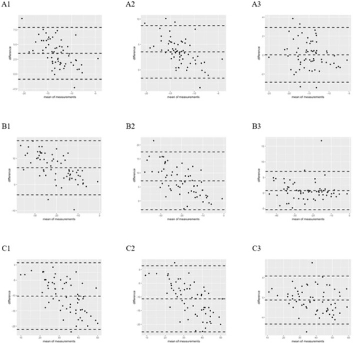
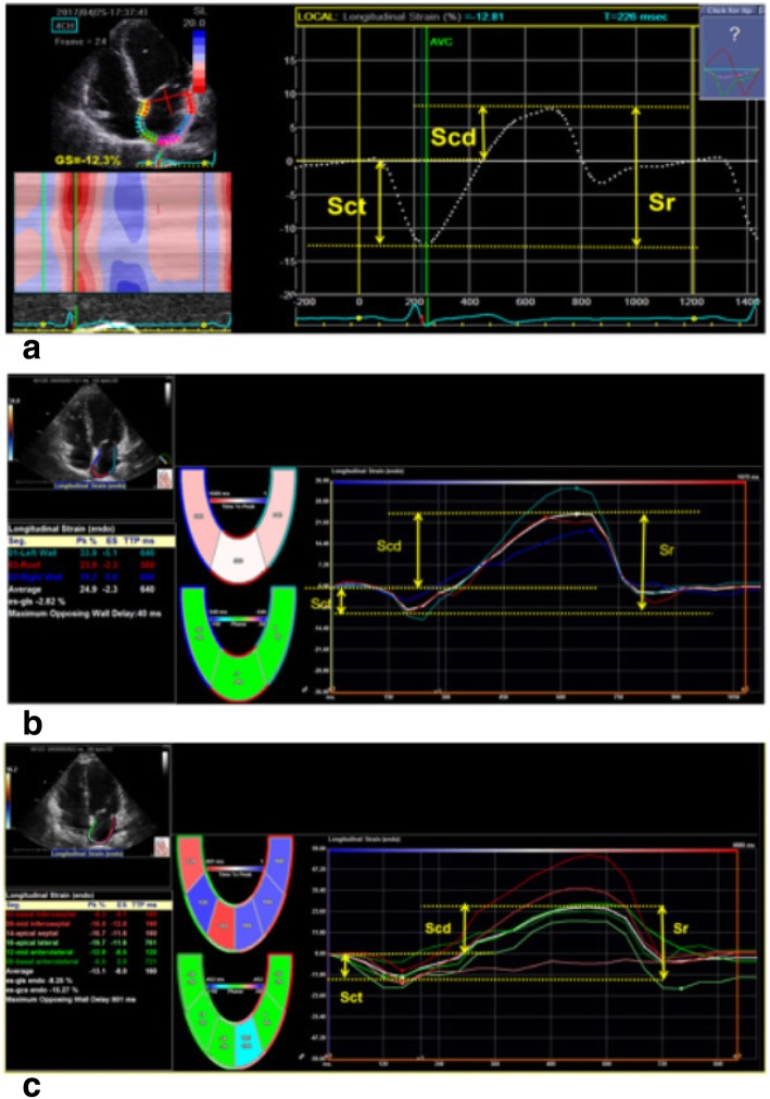
Sixty-four subjects (mean age: 44 ± 16 years, 50% males) underwent conventional echocardiograms using a GE Vivid 9 (GE Ultrasound, Horten, Norway) cardiac ultrasound system. Standard apical 4 and 2 chamber views of the left atrium were obtained in each subject with a frame-rate range of 40-71 frames/s. LA strain during the contraction phase (Sct), conduit phase (Scd), reservoir phase (Sr = Sct + Scd) were analyzed by 2 independent observers and 3 different software.
Sct, Scd, Sr were, respectively, - 11.26 ± 2.45%, - 16.77 ± 7.06%, and 28.03 ± 7.58% with VSS, - 14.77 ± 3.59%, - 23.17 ± 10.33%, and 38.23 ± 10.99% with VIS, and - 14.80 ± 3.88%, - 23.94 ± 10.48%, and 38.73 ± 11.56% when VIS was used. A comparison of strain measurements between VSS and VIS (VIS and VIS) showed VIS had significantly smaller mean differences and narrower limits of agreement. Similar results were observed in the coefficient of variation (CV) for measurements between VSS and VIS (VIS and VIS). Comparison of the intra-class correlation coefficients (ICCs) indicated that measurement reliability was weaker with VSS (ICC < 0.6) than with VIS (VIS and VIS) (ICC > 0.9). For intra-observer ICCs, VIS > VSS = VIS. For inter-observer ICCs, VSS > VIS > VIS.
Software measurement results of LA strain vary considerably. We recommended not measuring LA strain across vendor platforms.
二维斑点追踪超声心动图(2D-STE)能够通过心肌应变分析对左心房(LA)变形进行客观评估,心肌应变可通过不同的斑点追踪软件进行测量。本研究的目的是比较3种不同的市售软件的一致性,其中包括用于测量左心室的特定厂商软件(VSS)、用于测量左心室应变的独立于厂商的软件包(VIS)以及用于测量左心房应变的独立于厂商的软件包(VIS)。
64名受试者(平均年龄:44±16岁,50%为男性)使用GE Vivid 9(GE超声,挪威霍滕)心脏超声系统接受常规超声心动图检查。在每位受试者中获取标准的左心房心尖四腔和两腔视图,帧率范围为40-71帧/秒。由2名独立观察者和3种不同软件分析收缩期(Sct)、管道期(Scd)、储备期(Sr = Sct + Scd)的左心房应变。
使用VSS时,Sct、Scd、Sr分别为-11.26±2.45%、-16.77±7.06%和28.03±7.58%;使用VIS时,分别为-14.77±3.59%、-23.17±10.33%和38.23±10.99%;使用VIS时,分别为-14.80±3.88%、-23.94±10.48%和38.73±11.56%。VSS与VIS(VIS与VIS)之间的应变测量比较显示,VIS的平均差异显著更小,一致性界限更窄。在VSS与VIS(VIS与VIS)测量的变异系数(CV)中也观察到类似结果。组内相关系数(ICC)的比较表明,VSS(ICC < 0.6)的测量可靠性低于VIS(VIS与VIS)(ICC > 0.9)。对于观察者内ICC,VIS > VSS = VIS。对于观察者间ICC,VSS > VIS > VIS。
左心房应变软件测量结果差异很大。我们建议不要跨厂商平台测量左心房应变。