• 文献检索
  • 文档翻译
  • 深度研究
  • 学术资讯
  • Suppr Zotero 插件Zotero 插件
  • 邀请有礼
  • 套餐&价格
  • 历史记录
应用&插件
Suppr Zotero 插件Zotero 插件浏览器插件Mac 客户端Windows 客户端微信小程序
定价
高级版会员购买积分包购买API积分包
服务
文献检索文档翻译深度研究API 文档MCP 服务
关于我们
关于 Suppr公司介绍联系我们用户协议隐私条款
关注我们

Suppr 超能文献

核心技术专利:CN118964589B侵权必究
粤ICP备2023148730 号-1Suppr @ 2026

文献检索

告别复杂PubMed语法,用中文像聊天一样搜索,搜遍4000万医学文献。AI智能推荐,让科研检索更轻松。

立即免费搜索

文件翻译

保留排版,准确专业,支持PDF/Word/PPT等文件格式,支持 12+语言互译。

免费翻译文档

深度研究

AI帮你快速写综述,25分钟生成高质量综述,智能提取关键信息,辅助科研写作。

立即免费体验

采用图像分析方法测量角膜-翼状胬肉总面积。

Corneo-pterygium total area measurements utilising image analysis method.

作者信息

Mohd Radzi Hilmi, Khairidzan Mohd Kamal, Mohd Zulfaezal Che Amin, Azrin Esmady Ariffin

机构信息

Department of Optometry and Vision Science, Kulliyyah of Allied Health Sciences, International Islamic University Malaysia (IIUM), Kuantan, Pahang, Malaysia; Department of Ophthalmology, Kulliyyah of Medicine, International Islamic University Malaysia (IIUM), Kuantan, Pahang, Malaysia.

Department of Ophthalmology, Kulliyyah of Medicine, International Islamic University Malaysia (IIUM), Kuantan, Pahang, Malaysia.

出版信息

J Optom. 2019 Oct-Dec;12(4):272-277. doi: 10.1016/j.optom.2019.04.001. Epub 2019 May 13.

DOI:10.1016/j.optom.2019.04.001
PMID:31097348
原文链接:https://pmc.ncbi.nlm.nih.gov/articles/PMC6978598/
Abstract

PURPOSE

To describe an objective method to accurately quantify corneo-pterygium total area (CPTA) by utilising image analysis method and to evaluate its association with corneal astigmatism (CA).

METHODS

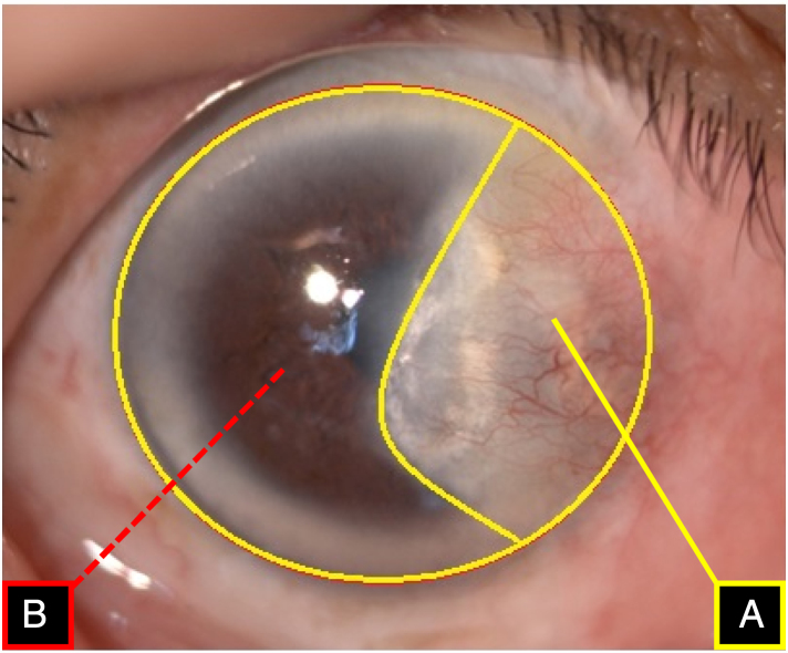
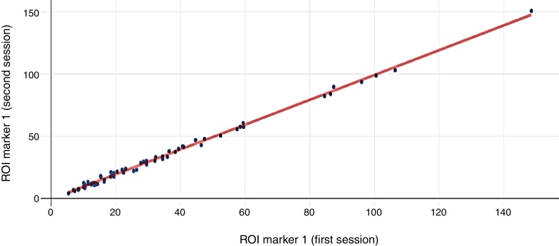
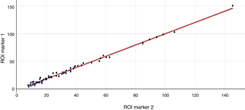
120 primary pterygium participants were selected from patients who visited an ophthalmology clinic. We adopted image analysis software in calculating the size of invading pterygium to the cornea. The marking of the calculated area was done manually, and the total area size was measured in pixel. The computed area is defined as the area from the apex of pterygium to the limbal-corneal border. Then, from the pixel, it was transformed into a percentage (%), which represents the CPTA relative to the entire corneal surface area. Intra- and inter-observer reliability testing were performed by repeating the tracing process twice with a different sequence of images at least one (1) month apart. Intraclass correlation (ICC) and scatter plot were used to describe the reliability of measurement.

RESULTS

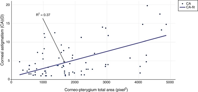
The overall mean (N=120) of CPTA was 45.26±13.51% (CI: 42.38-48.36). Reliability for region of interest (ROI) demarcation of CPTA were excellent with intra and inter-agreement of 0.995 (95% CI, 0.994-0.998; P<0.001) and 0.994 (95% CI, 0.992-0.997; P<0.001) respectively. The new method was positively associated with corneal astigmatism (P<0.01). This method was able to predict 37% of the variance in CA compared to 21% using standard method.

CONCLUSIONS

Image analysis method is useful, reliable and practical in the clinical setting to objectively quantify actual pterygium size, shapes and its effects on the anterior corneal curvature.

摘要

目的

描述一种利用图像分析方法准确量化角膜-翼状胬肉总面积(CPTA)的客观方法,并评估其与角膜散光(CA)的相关性。

方法

从眼科门诊就诊的患者中选取120例原发性翼状胬肉患者。我们采用图像分析软件计算翼状胬肉侵入角膜的大小。计算区域的标记通过手动完成,总面积大小以像素为单位进行测量。计算区域定义为从翼状胬肉顶端到角膜缘-角膜边界的区域。然后,从像素值转换为百分比(%),表示CPTA相对于整个角膜表面积的比例。通过至少间隔一(1)个月以不同图像序列重复追踪过程两次,进行观察者内和观察者间可靠性测试。组内相关系数(ICC)和散点图用于描述测量的可靠性。

结果

CPTA的总体平均值(N = 120)为45.26±13.51%(CI:42.38 - 48.36)。CPTA感兴趣区域(ROI)划分的可靠性极佳,观察者内一致性为0.995(95% CI,0.994 - 0.998;P < 0.001),观察者间一致性为0.994(95% CI,0.992 - 0.997;P < 0.001)。新方法与角膜散光呈正相关(P < 0.01)。与标准方法相比,该方法能够预测37%的角膜散光变异,而标准方法为21%。

结论

图像分析方法在临床环境中对于客观量化实际翼状胬肉大小、形状及其对角膜前曲率的影响是有用、可靠且实用的。

https://cdn.ncbi.nlm.nih.gov/pmc/blobs/33f7/6978598/cf0a88578683/gr4.jpg
https://cdn.ncbi.nlm.nih.gov/pmc/blobs/33f7/6978598/fe909a101fa5/gr1.jpg
https://cdn.ncbi.nlm.nih.gov/pmc/blobs/33f7/6978598/9ae966cdee1d/gr2.jpg
https://cdn.ncbi.nlm.nih.gov/pmc/blobs/33f7/6978598/b3a2dbc01010/gr3.jpg
https://cdn.ncbi.nlm.nih.gov/pmc/blobs/33f7/6978598/cf0a88578683/gr4.jpg
https://cdn.ncbi.nlm.nih.gov/pmc/blobs/33f7/6978598/fe909a101fa5/gr1.jpg
https://cdn.ncbi.nlm.nih.gov/pmc/blobs/33f7/6978598/9ae966cdee1d/gr2.jpg
https://cdn.ncbi.nlm.nih.gov/pmc/blobs/33f7/6978598/b3a2dbc01010/gr3.jpg
https://cdn.ncbi.nlm.nih.gov/pmc/blobs/33f7/6978598/cf0a88578683/gr4.jpg

相似文献

1
Corneo-pterygium total area measurements utilising image analysis method.采用图像分析方法测量角膜-翼状胬肉总面积。
J Optom. 2019 Oct-Dec;12(4):272-277. doi: 10.1016/j.optom.2019.04.001. Epub 2019 May 13.
2
Correlation between pterygium size and induced corneal astigmatism.翼状胬肉大小与诱导性角膜散光之间的相关性。
Cornea. 1998 Jan;17(1):28-30. doi: 10.1097/00003226-199801000-00005.
3
[Impact of pterygium size on corneal topography and visual acuity - a prospective clinical cross-sectional study].[翼状胬肉大小对角膜地形图和视力的影响——一项前瞻性临床横断面研究]
Klin Monbl Augenheilkd. 2001 Sep;218(9):609-15. doi: 10.1055/s-2001-17639.
4
Quantification of Astigmatism Induced by Pterygium Using Automated Image Analysis.使用自动图像分析技术对翼状胬肉所致散光进行定量分析。
Cornea. 2016 Mar;35(3):370-6. doi: 10.1097/ICO.0000000000000728.
5
[Quantitative evaluation of corneal irregular astigmatism using computed corneal topography].
Nippon Ganka Gakkai Zasshi. 1995 Mar;99(3):296-301.
6
Effects of fibrovascular traction and pooling of tears on corneal topographic changes induced by pterygium.纤维血管牵引和泪液积聚对翼状胬肉所致角膜地形变化的影响。
Eye (Lond). 2003 May;17(4):492-6. doi: 10.1038/sj.eye.6700377.
7
The effect of limbal autograft location at primary pterygium excision on anterior and posterior corneal astigmatism: a comparative prospective study.原发性翼状胬肉切除术中角膜缘移植物位置对前后角膜散光的影响:一项比较前瞻性研究。
Int Ophthalmol. 2022 Jul;42(7):2235-2243. doi: 10.1007/s10792-022-02223-7. Epub 2022 Jan 23.
8
Pterygium-induced corneal refractive changes.翼状胬肉引起的角膜屈光变化。
Indian J Ophthalmol. 2007 Sep-Oct;55(5):383-6. doi: 10.4103/0301-4738.33829.
9
Analysis of pterygium size and induced corneal astigmatism.翼状胬肉大小及诱导性角膜散光分析。
Cornea. 2008 May;27(4):434-8. doi: 10.1097/ICO.0b013e3181656448.
10
A comparative study of preoperative and postoperative changes in corneal astigmatism after pterygium excision by different techniques.不同手术方式切除翼状胬肉术后角膜散光的变化比较
Indian J Ophthalmol. 2019 Jul;67(7):1036-1039. doi: 10.4103/ijo.IJO_1921_18.

引用本文的文献

1
Safety and efficacy of "hydro-fluorescein" technique in removing Tenon in pterygium surgery: a 1-year follow-up study.“氢荧光素”技术在翼状胬肉手术中切除Tenon囊的安全性和有效性:一项为期1年的随访研究。
Eye (Lond). 2025 Apr;39(6):1081-1085. doi: 10.1038/s41433-024-03539-7. Epub 2024 Dec 10.
2
Artificial intelligence assisted pterygium diagnosis: current status and perspectives.人工智能辅助翼状胬肉诊断:现状与展望
Int J Ophthalmol. 2023 Sep 18;16(9):1386-1394. doi: 10.18240/ijo.2023.09.04. eCollection 2023.
3
Advances in artificial intelligence applications for ocular surface diseases diagnosis.

本文引用的文献

1
Prediction of Changes in Visual Acuity and Contrast Sensitivity Function by Tissue Redness after Pterygium Surgery.翼状胬肉切除术后组织发红对视力和对比敏感度功能变化的预测
Curr Eye Res. 2017 Jun;42(6):852-856. doi: 10.1080/02713683.2016.1250277. Epub 2017 Jan 24.
2
Risk Factors for Recurrence After Pterygium Surgery: An Image Analysis Study.翼状胬肉手术后复发的危险因素:一项图像分析研究。
Cornea. 2016 Aug;35(8):1097-103. doi: 10.1097/ICO.0000000000000853.
3
Quantification of Astigmatism Induced by Pterygium Using Automated Image Analysis.
人工智能在眼表疾病诊断中的应用进展。
Front Cell Dev Biol. 2022 Dec 20;10:1107689. doi: 10.3389/fcell.2022.1107689. eCollection 2022.
4
Comparison of Efficacy and Safety of Two Commercial Artificial Tears Between 0.18% and 0.3% Sodium Hyaluronate for Corneal Epithelial Healing in Pterygium Excision with Conjunctival Autograft Transplantation: A Study Protocol for a Randomized Controlled Trial.0.18%至0.3%透明质酸钠的两种市售人工泪液在翼状胬肉切除联合自体结膜移植术中促进角膜上皮愈合的疗效与安全性比较:一项随机对照试验的研究方案
Clin Ophthalmol. 2022 Nov 30;16:3935-3944. doi: 10.2147/OPTH.S388276. eCollection 2022.
5
Computer-Assisted Pterygium Screening System: A Review.计算机辅助翼状胬肉筛查系统:综述
Diagnostics (Basel). 2022 Mar 5;12(3):639. doi: 10.3390/diagnostics12030639.
6
A Novel System for Measuring Pterygium's Progress Using Deep Learning.一种使用深度学习测量翼状胬肉进展的新型系统。
Front Med (Lausanne). 2022 Feb 14;9:819971. doi: 10.3389/fmed.2022.819971. eCollection 2022.
7
Group and Shuffle Convolutional Neural Networks with Pyramid Pooling Module for Automated Pterygium Segmentation.基于金字塔池化模块的分组与随机卷积神经网络用于翼状胬肉自动分割
Diagnostics (Basel). 2021 Jun 17;11(6):1104. doi: 10.3390/diagnostics11061104.
使用自动图像分析技术对翼状胬肉所致散光进行定量分析。
Cornea. 2016 Mar;35(3):370-6. doi: 10.1097/ICO.0000000000000728.
4
Influence of Pterygium on Corneal Biomechanical Properties.翼状胬肉对角膜生物力学特性的影响。
Curr Eye Res. 2016 Jul;41(7):913-6. doi: 10.3109/02713683.2015.1080281. Epub 2015 Oct 16.
5
Astigmatic changes following pterygium removal: comparison of 5 different methods.翼状胬肉切除术后的散光变化:5 种不同方法的比较。
Indian J Ophthalmol. 2013 Mar;61(3):104-8. doi: 10.4103/0301-4738.109379.
6
Effects of pterygium surgery on front and back corneal surfaces and anterior segment parameters.翼状胬肉手术对角膜前后表面及眼前节参数的影响。
Int Ophthalmol. 2012 Jun;32(3):251-7. doi: 10.1007/s10792-012-9560-2. Epub 2012 Apr 7.
7
Effect of pterygia on refractive indices, corneal topography, and ocular aberrations.翼状胬肉对屈光度、角膜地形和眼像差的影响。
Cornea. 2011 Jan;30(1):24-9. doi: 10.1097/ICO.0b013e3181dc814e.
8
Objective grading of the anterior eye.眼前部的客观分级。
Optom Vis Sci. 2009 Mar;86(3):273-8. doi: 10.1097/OPX.0b013e3181981976.
9
Analysis of pterygium size and induced corneal astigmatism.翼状胬肉大小及诱导性角膜散光分析。
Cornea. 2008 May;27(4):434-8. doi: 10.1097/ICO.0b013e3181656448.
10
The use of fractal analysis and photometry to estimate the accuracy of bulbar redness grading scales.使用分形分析和光度测定法评估球结膜充血分级量表的准确性。
Invest Ophthalmol Vis Sci. 2008 Apr;49(4):1398-406. doi: 10.1167/iovs.07-1306.