Wu Chi-Chuan, Yeow Kee-Min, Yeow Yun-Jen
Chang Gung Memorial Hospital, Chang Gung University, Department of Orthopedic Surgery, Taipei, Taiwan.
Chang Gung Memorial Hospital, Chang Gung University, Department of Imaging Diagnosis, Taipei, Taiwan.
Acta Orthop Traumatol Turc. 2019 Jul;53(4):287-291. doi: 10.1016/j.aott.2019.04.011. Epub 2019 May 15.
The aim of this study was to evaluate the varied influence of femoral or tibial component on Quadriceps angles (Q-angle) measured with magnetic resonance image (MRI) and full-length standing scanogram (FLSS) techniques.
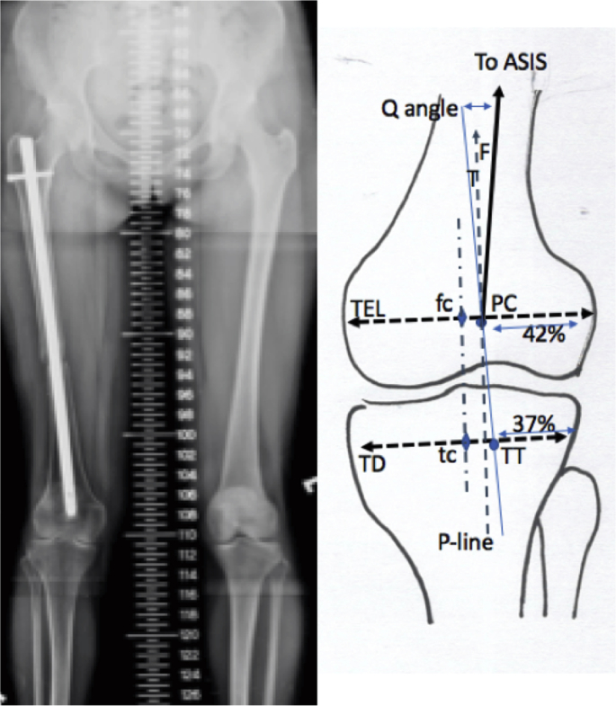
Two groups of patients were studied. The first group underwent MRI studies and the second group underwent FLSS studies. Two-step procedures were carried out. Knee MRI in 60 consecutive adult patients simply taken for meniscus or ligament injuries were utilized at the first step. The standardized patellar center (PC) and tibial tubercle (TT) on the frontal plane of MRI were positioned. At the second step, the FLSS in other 100 consecutive young adult patients taken for chronic unilateral lower extremity injuries were used for locating the two landmarks from MRI. The Q-angle was then determined on the anterior superior iliac spine, standardized PC, and TT on the FLSS.
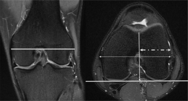
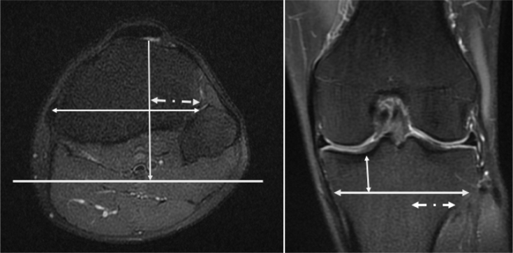
For 60 patients, the standardized PC was at the point 42% from the lateral end of the trans-epicondylar line of the femur. The TT was at the point 2 cm distal to the tibial articular surface and 37% from the lateral end of the tibial width. For 100 patients, the Q-angle was an average of 9.5° and 65.2% of the Q-angle was contributed by the upper arm (the femur). Women had a larger Q-angle (10.1° vs. 8.8°, p = 0.02) and a shorter femur (41.1 vs. 44.7 cm, p < 0.001).
The Q-angle is about 9.5° with 65.2% contributed by the femur. The Q-angle may mainly be influenced by the femoral component.
Level IV, Diagnostic Study.
本研究旨在评估股骨或胫骨组件对采用磁共振成像(MRI)和全长站立扫描图(FLSS)技术测量的股四头肌角(Q角)的不同影响。
对两组患者进行了研究。第一组接受MRI检查,第二组接受FLSS检查。进行了两步操作。第一步,利用60例单纯因半月板或韧带损伤而接受检查的连续成年患者的膝关节MRI。在MRI的额平面上定位标准化髌骨中心(PC)和胫骨结节(TT)。第二步,将另外100例因慢性单侧下肢损伤而接受检查的连续年轻成年患者的FLSS用于从MRI中定位这两个标志点。然后在FLSS上的髂前上棘、标准化PC和TT处测定Q角。
对于60例患者,标准化PC位于距股骨髁上连线外侧端42%处。TT位于胫骨关节面远端2 cm处,距胫骨宽度外侧端37%处。对于100例患者,Q角平均为9.5°,其中65.2%由上臂(股骨)贡献。女性的Q角更大(10.1°对8.8°,p = 0.02),股骨更短(41.1对44.7 cm,p < 0.001)。
Q角约为9.5°,其中65.2%由股骨贡献。Q角可能主要受股骨组件的影响。
IV级,诊断性研究。