Tanner Jared J, Amin Manish, Hardcastle Cheshire, Parvataneni Hari, Vaillancourt David E, Mareci Thomas H, Price Catherine C
Department of Clinical and Health Psychology, University of Florida, Gainesville, FL, United States.
Department of Physics, University of Florida, Gainesville, FL, United States.
Front Aging Neurosci. 2019 May 16;11:117. doi: 10.3389/fnagi.2019.00117. eCollection 2019.
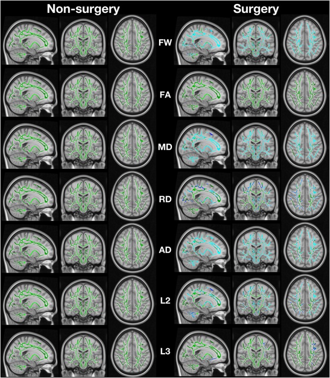
For adults age 65 and older, the brain shows acute functional connectivity decreases after total knee arthroplasty with the severity of change predicted by preoperative cognitive function and brain disease burden. The extent of acute structural microstructural brain changes acutely after surgery remains unknown within the literature. For the current study, we report on the severity of acute post-surgery microstructural brain changes as measured by diffusion imaging and free-water analysis. Participants who underwent total knee arthroplasty under general anesthesia and non-surgery peers were part of a federally funded prospective cohort investigation involving participants. Recruitment occurred between 2013 and 2017. Data were collected in outpatient and inpatient settings within a university-affiliated medical center. A total of 232 TKA patients were referred by the study surgeon and contacted for study inclusion. Of these, 78 met inclusion and exclusion criteria and completed assessment. Five participants were excluded due to anesthetic protocol changes (spinal instead of general) with an additional 12 excluded for imaging-related complications. The total included sample size was 61. A total of 127 non-surgery participants were screened with 66 enrolled. One non-surgery participant was excluded for an imaging-related complication. Total knee arthroplasty and general anesthetic protocols were standardized. Participants received preoperative neurocognitive assessment and brain magnetic resonance imaging, with repeat imaging 48 h after surgery or pseudo surgery. Free-water analyses were performed using diffusion weighted images and tract-based spatial statistics with baseline cognitive data used to predict free-water changes. Surgery participants had widespread increases in white matter free-water. Surgery participants with higher cognitive functions as measured by immediate memory and less evidence of brain atrophy and disease (i.e., brain integrity) had greater free-water increase. Non-surgery peers had no free-water change. We interpret the surgery group's free-water change as indicating widespread brain white matter glial response, with greater change indicative of better brain response to the acute surgery/anesthesia experience.
对于65岁及以上的成年人,全膝关节置换术后大脑会出现急性功能连接性下降,其变化的严重程度可由术前认知功能和脑部疾病负担预测。术后大脑急性结构微观结构变化的程度在文献中尚不清楚。在本研究中,我们报告了通过扩散成像和自由水分析测量的术后急性微观结构大脑变化的严重程度。接受全身麻醉下全膝关节置换术的参与者和非手术对照组是一项由联邦政府资助的前瞻性队列研究的一部分。招募工作于2013年至2017年期间进行。数据在大学附属医疗中心的门诊和住院环境中收集。共有232名全膝关节置换术患者由研究外科医生转诊并被联系以纳入研究。其中,78名符合纳入和排除标准并完成评估。5名参与者因麻醉方案改变(从全身麻醉改为脊髓麻醉)被排除,另有12名因与成像相关的并发症被排除。纳入的样本总数为61名。共筛选了127名非手术参与者,其中66名被纳入。1名非手术参与者因与成像相关的并发症被排除。全膝关节置换术和全身麻醉方案均标准化。参与者接受术前神经认知评估和脑部磁共振成像,并在手术后或假手术后48小时进行重复成像。使用扩散加权图像和基于束的空间统计学进行自由水分析,并使用基线认知数据来预测自由水变化。手术参与者的白质自由水普遍增加。通过即时记忆测量具有较高认知功能且脑萎缩和疾病证据较少(即脑完整性较好)的手术参与者,其自由水增加幅度更大。非手术对照组的自由水没有变化。我们将手术组的自由水变化解释为表明广泛的脑白质胶质细胞反应,变化越大表明大脑对急性手术/麻醉经历的反应越好。