Department of Biomedicine & Health Sciences, Graduate School, The Catholic University of Korea, Seoul, Republic of Korea.
Departments of Biomedical Engineering, Konyang University, Daejeon, Republic of Korea.
J Exp Clin Cancer Res. 2019 Jun 14;38(1):258. doi: 10.1186/s13046-019-1225-9.
Pancreatic ductal adenocarcinoma (PDAC) is a stroma-rich carcinoma, and pancreatic stellate cells (PSCs) are a major component of this dense stroma. PSCs play significant roles in metastatic progression and chemoresistance through cross-talk with cancer cells. Preclinical in vitro tumor model of invasive phenotype should incorporate three-dimensional (3D) culture of cancer cells and PSCs in extracellular matrix (ECM) for clinical relevance and predictability.
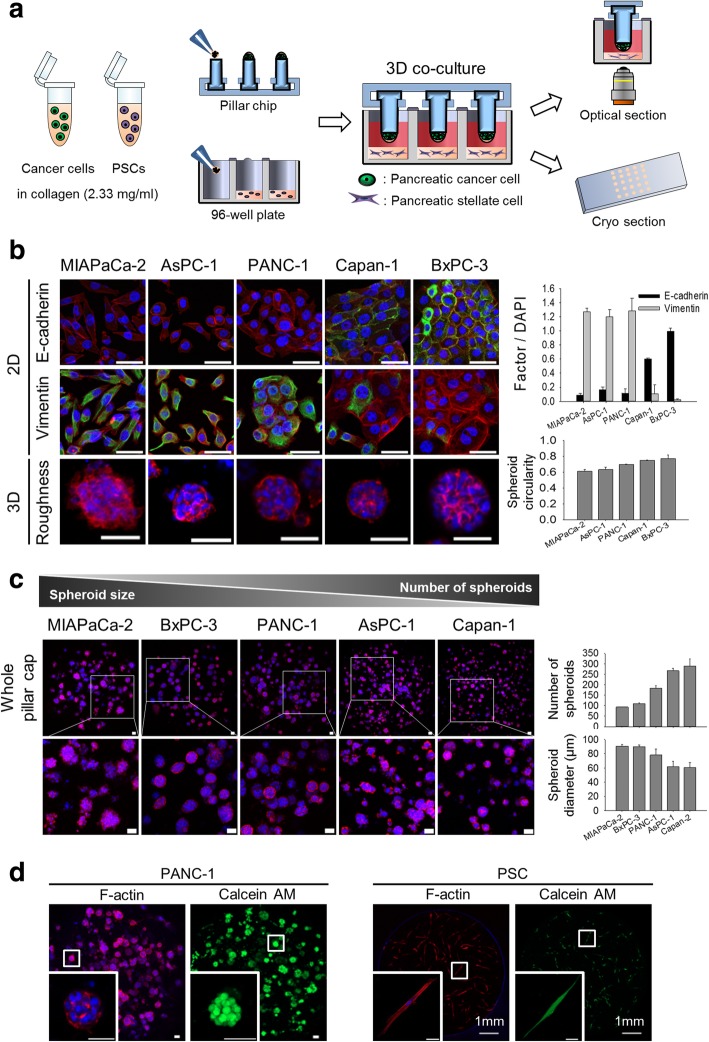
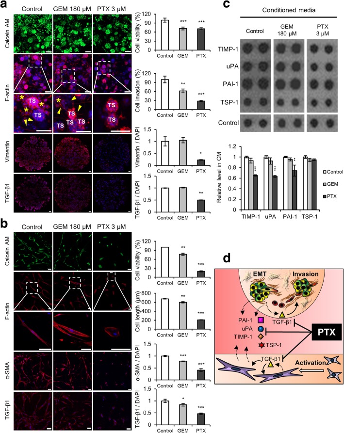
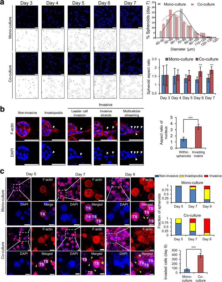
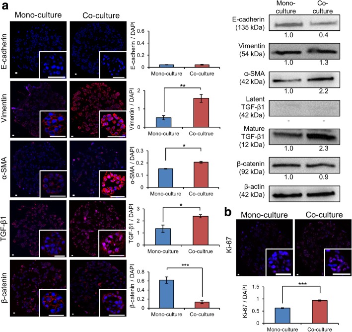
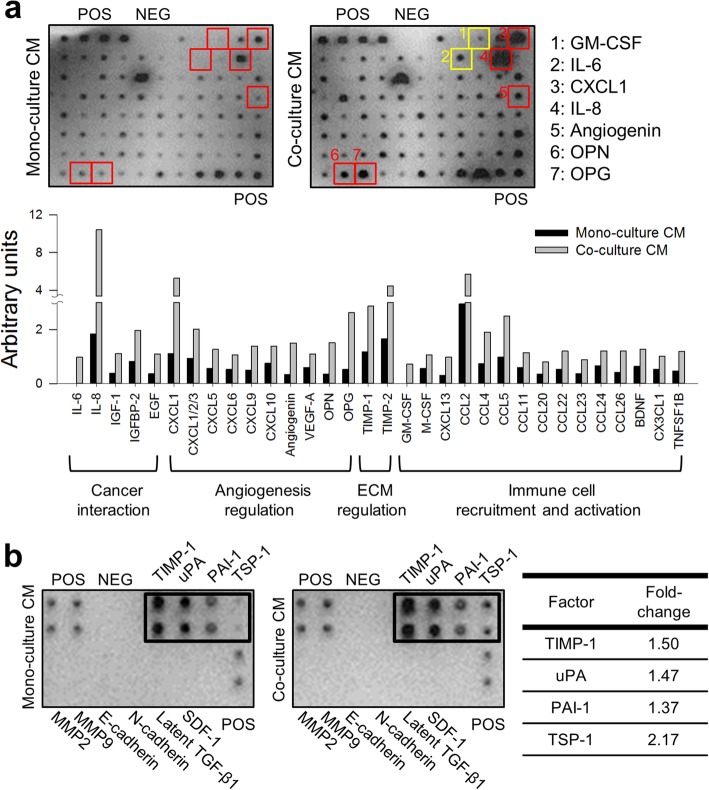
PANC-1 cells were cultured as tumor spheroids (TSs) using our previously developed minipillar chips, and co-cultured with PSCs, both embedded in collagen gels. Effects of PSC co-culture on ECM fiber network, invasive migration of cancer cells, and expression of epithelial-mesenchymal transition (EMT)-related proteins were examined. Conditioned media was also analyzed for secreted factors involved in cancer cell-PSC interactions. Inhibitory effect on cancer cell invasion was compared between gemcitabine and paclitaxel at an equitoxic concentration in PANC-1 TSs co-cultured with PSCs.
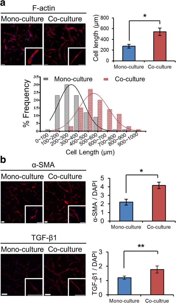
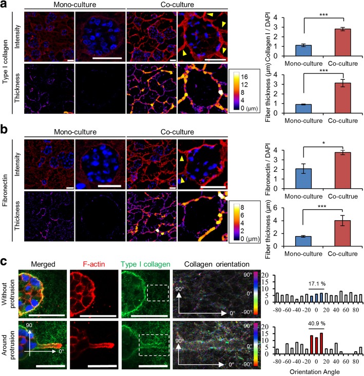
Co-culture condition was optimized for the growth of TSs, activation of PSCs, and their interaction. Increase in cancer cell invasion via ECM remodeling, invadopodia formation and EMT, as well as drug resistance was recapitulated in the TS-PSC co-culture, and appeared to be mediated by cancer cell-PSC interaction via multiple secreted factors, including IL-6, IL-8, IGF-1, EGF, TIMP-1, uPA, PAI-1, and TSP-1. Compared to gemcitabine, paclitaxel showed a greater anti-invasive activity, which was attributed to suppresion of invadopodia formation in cancer cells as well as to PSC-specific cytotoxicity abrogating its paracrine signaling.
Here, we established 3D co-culture of TSs of PANC-1 cells and PSCs using minipillar histochips as a novel tumoroid model of PDAC. Our results indicate usefulness of the present co-culture model and multiplex quantitative analysis method not only in studying the role of PSCs and their interactions with tumor cell towards metastatic progression, but also in the drug evaluation of stroma-targeting drugs.
胰腺导管腺癌(PDAC)是一种富含基质的癌,胰腺星状细胞(PSC)是这种致密基质的主要组成部分。PSC 通过与癌细胞的相互作用,在转移进展和化疗耐药性方面发挥重要作用。临床相关性和可预测性要求将癌症细胞和 PSC 三维(3D)培养在细胞外基质(ECM)中,建立临床前体外侵袭表型肿瘤模型。
使用我们之前开发的微柱芯片培养 PANC-1 细胞作为肿瘤球体(TS),并将其与嵌入胶原凝胶中的 PSC 共培养。研究 PSC 共培养对 ECM 纤维网络、癌细胞侵袭性迁移以及上皮-间充质转化(EMT)相关蛋白表达的影响。还分析了条件培养基中涉及癌细胞-PSC 相互作用的分泌因子。在与 PSC 共培养的 PANC-1 TS 中,比较了吉西他滨和紫杉醇在等毒性浓度下对癌细胞侵袭的抑制作用。
优化了 TS 生长、PSC 激活及其相互作用的共培养条件。在 TS-PSC 共培养中,通过 ECM 重塑、侵袭伪足形成和 EMT 以及药物耐药性,重现了癌细胞侵袭的增加,并且似乎是通过多种分泌因子介导的,包括 IL-6、IL-8、IGF-1、EGF、TIMP-1、uPA、PAI-1 和 TSP-1。与吉西他滨相比,紫杉醇显示出更强的抗侵袭活性,这归因于抑制癌细胞侵袭伪足形成以及 PSC 特异性细胞毒性,从而阻断其旁分泌信号。
在这里,我们使用微柱组织芯片建立了 PANC-1 细胞 TS 和 PSC 的 3D 共培养,作为 PDAC 的新型肿瘤模型。我们的结果表明,这种共培养模型和多重定量分析方法不仅可用于研究 PSC 及其与肿瘤细胞相互作用在转移进展方面的作用,还可用于评估针对基质的药物。