文献检索文档翻译深度研究
Suppr Zotero 插件Zotero 插件
邀请有礼套餐&价格历史记录

新学期,新优惠

限时优惠:9月1日-9月22日

30天高级会员仅需29元

1天体验卡首发特惠仅需5.99元

了解详情
不再提醒
插件&应用
Suppr Zotero 插件Zotero 插件浏览器插件Mac 客户端Windows 客户端微信小程序
高级版
套餐订阅购买积分包
AI 工具
文献检索文档翻译深度研究
关于我们
关于 Suppr公司介绍联系我们用户协议隐私条款
关注我们

Suppr 超能文献

核心技术专利:CN118964589B侵权必究
粤ICP备2023148730 号-1Suppr @ 2025

用不同类别的活细胞染料对人骨髓间充质干细胞进行标记:稳健性和毒性。

Labeling of human mesenchymal stem cells with different classes of vital stains: robustness and toxicity.

机构信息

NeuroRepair Department, Mossakowski Medical Research Centre, Polish Academy of Sciences, Warsaw, Poland.

Division of MR Research, Russell H. Morgan Department of Radiology and Radiological Science, The Johns Hopkins University School of Medicine, Baltimore, USA.

出版信息

Stem Cell Res Ther. 2019 Jun 25;10(1):187. doi: 10.1186/s13287-019-1296-8.


DOI:10.1186/s13287-019-1296-8
PMID:31238982
原文链接:https://pmc.ncbi.nlm.nih.gov/articles/PMC6593614/
Abstract

BACKGROUND: Mesenchymal stem cell (MSC) transplantation has been explored as a new clinical approach to repair injured tissues. However, in order to evaluate the therapeutic activity of MSC, cell tracking techniques are required to determine the fate of transplanted cells in both preclinical and clinical studies. In these aspects, different vital stains offer the potential for labeling and monitoring of grafted cells in vivo. It is desirable to have tracking agents which have long-term stability, are not toxic to the cells, and do not affect cell function. METHODS: Here, we selected three different labels: CellTracker™ Green CMFDA, eGFP-mRNA (genetic pre-tag), and Molday ION Rhodamine B™ (nanoparticle-based fluorescent and magnetic label) and performed extensive analysis of their influence on in vitro expansion of human bone marrow-derived mesenchymal stem cells (hBM-MSCs), as well as potential of affecting therapeutic activity and the impact on the durability of staining. RESULTS: 
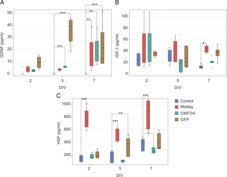
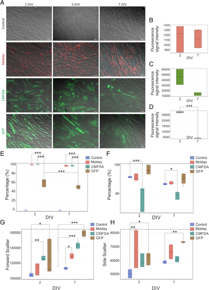
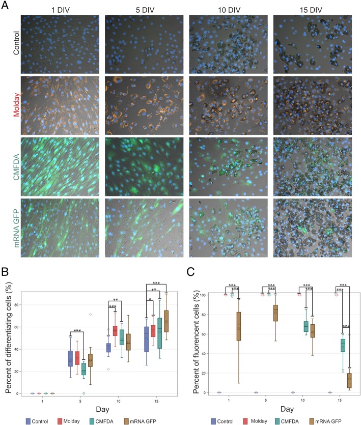
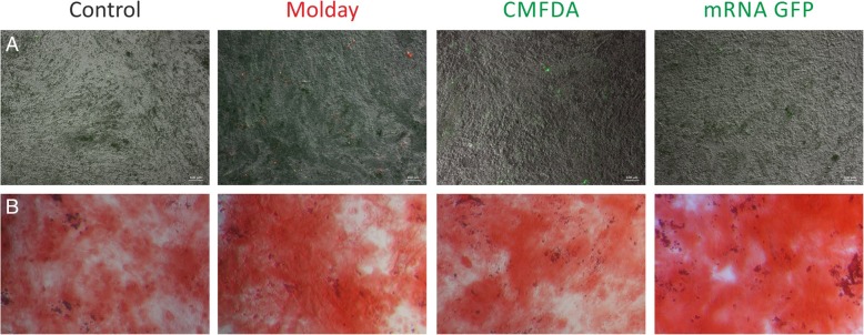
Our study showed that basic hBM-MSC characteristics and functions might be affected by labeling. We observed strong alterations of metabolic activity and morphology after eGFP and CellTracker™ Green CMFDA hBM-MSC staining. Molday ION Rhodamine B™ labeling revealed superior properties relatively to other vital stains. The relative expression level of most of the investigated growth factors remained stable after cell labeling, but we have observed some changes in the case of EGF, GDNF, HGF, and IGF gene expression. CONCLUSIONS: Taken together, we suggest performing similar to ours extensive analysis prior to using any cell label to tag MSC in experiments, as it can thoroughly bias results.

摘要

背景:间充质干细胞(MSC)移植已被探索作为修复受损组织的一种新的临床方法。然而,为了评估 MSC 的治疗活性,需要细胞跟踪技术来确定临床前和临床研究中移植细胞的命运。在这些方面,不同的活体染料为体内标记和监测移植细胞提供了潜力。理想的跟踪剂应具有长期稳定性、对细胞无毒、且不影响细胞功能。

方法:在这里,我们选择了三种不同的标记物:CellTracker™ Green CMFDA、eGFP-mRNA(遗传预标记)和 Molday ION Rhodamine B™(基于纳米颗粒的荧光和磁性标记),并对它们对人骨髓间充质干细胞(hBM-MSCs)体外扩增的影响进行了广泛分析,以及对治疗活性的潜在影响和对染色耐久性的影响。

结果:我们的研究表明,基本的 hBM-MSC 特征和功能可能会受到标记的影响。我们观察到 eGFP 和 CellTracker™ Green CMFDA hBM-MSC 染色后代谢活性和形态发生强烈改变。Molday ION Rhodamine B™ 标记与其他活体染料相比具有优越的特性。在细胞标记后,大多数研究的生长因子的相对表达水平保持稳定,但我们观察到在 EGF、GDNF、HGF 和 IGF 基因表达的情况下发生了一些变化。

结论:综上所述,我们建议在实验中使用任何细胞标记物标记 MSC 之前,像我们这样进行广泛的分析,因为它会严重影响结果。

https://cdn.ncbi.nlm.nih.gov/pmc/blobs/818a/6593614/3019d79cbb10/13287_2019_1296_Fig8_HTML.jpg
https://cdn.ncbi.nlm.nih.gov/pmc/blobs/818a/6593614/ed36239e2e19/13287_2019_1296_Fig1_HTML.jpg
https://cdn.ncbi.nlm.nih.gov/pmc/blobs/818a/6593614/eb0f8b3e17b6/13287_2019_1296_Fig2_HTML.jpg
https://cdn.ncbi.nlm.nih.gov/pmc/blobs/818a/6593614/29c12f301d3f/13287_2019_1296_Fig3_HTML.jpg
https://cdn.ncbi.nlm.nih.gov/pmc/blobs/818a/6593614/e51766d4420d/13287_2019_1296_Fig4_HTML.jpg
https://cdn.ncbi.nlm.nih.gov/pmc/blobs/818a/6593614/0494d4eb5901/13287_2019_1296_Fig5_HTML.jpg
https://cdn.ncbi.nlm.nih.gov/pmc/blobs/818a/6593614/99d27bbb1c2f/13287_2019_1296_Fig6_HTML.jpg
https://cdn.ncbi.nlm.nih.gov/pmc/blobs/818a/6593614/2d6aedc0488d/13287_2019_1296_Fig7_HTML.jpg
https://cdn.ncbi.nlm.nih.gov/pmc/blobs/818a/6593614/3019d79cbb10/13287_2019_1296_Fig8_HTML.jpg

相似文献

[1]
Labeling of human mesenchymal stem cells with different classes of vital stains: robustness and toxicity.

Stem Cell Res Ther. 2019-6-25

[2]
Toward an ideal animal model to trace donor cell fates after stem cell therapy: production of stably labeled multipotent mesenchymal stem cells from bone marrow of transgenic pigs harboring enhanced green fluorescence protein gene.

J Anim Sci. 2011-6-24

[3]
Mesenchymal stem cell labeling and in vitro MR characterization at 1.5 T of new SPIO contrast agent: Molday ION Rhodamine-B™.

Contrast Media Mol Imaging. 2010-8-5

[4]
Imaging of extracellular vesicles derived from human bone marrow mesenchymal stem cells using fluorescent and magnetic labels.

Int J Nanomedicine. 2018-3-19

[5]
Tissue source determines the differentiation potentials of mesenchymal stem cells: a comparative study of human mesenchymal stem cells from bone marrow and adipose tissue.

Stem Cell Res Ther. 2017-12-6

[6]
Transplantation of MSCs Overexpressing HGF into a Rat Model of Liver Fibrosis.

Mol Imaging Biol. 2016-2

[7]
Impact of c-MYC expression on proliferation, differentiation, and risk of neoplastic transformation of human mesenchymal stromal cells.

Stem Cell Res Ther. 2019-3-5

[8]
In vivo tracking of novel SPIO-Molday ION rhodamine-B™-labeled human bone marrow-derived mesenchymal stem cells after lentivirus- mediated COX-2 silencing: a preliminary study.

Curr Gene Ther. 2014

[9]
Long-term detection of fluorescently labeled human mesenchymal stem cell in vitro and in vivo by semi-automated microscopy.

Tissue Eng Part C Methods. 2011-12-13

[10]
Mesenchymal stem cells derived from human induced pluripotent stem cells retain adequate osteogenicity and chondrogenicity but less adipogenicity.

Stem Cell Res Ther. 2015-8-18

引用本文的文献

[1]
Smart CAR-T Nanosymbionts: archetypes and proto-models.

Front Immunol. 2025-8-12

[2]
Neuro-Cells Mitigate Amyloid Plaque Formation and Behavioral Deficits in the APPswe/PS1dE9 Model of Alzheimer Disease While Also Reducing IL-6 Production in Human Monocytes.

Cells. 2025-7-29

[3]
Dielectrophoretic Microfluidic Designs for Precision Cell Enrichments and Highly Viable Label-Free Bacteria Recovery from Blood.

Micromachines (Basel). 2025-2-19

[4]
Biodistribution and persistence of human umbilical cord-derived mesenchymal stem cells in NCG mice: a comparative study.

Future Sci OA. 2025-12

[5]
Label-free ghost cytometry for manufacturing of cell therapy products.

Sci Rep. 2024-9-19

[6]
Fluorescence-Based Mono- and Multimodal Imaging for In Vivo Tracking of Mesenchymal Stem Cells.

Biomolecules. 2023-12-13

[7]
Preclinical Short-term and Long-term Safety of Human Bone Marrow Mesenchymal Stem Cells.

Cell Transplant. 2023

[8]
Virtual staining for pixel-wise and quantitative analysis of single cell images.

Sci Rep. 2023-11-6

[9]
Mesenchymal stem cell engineering by ARCA analog-capped mRNA.

Mol Ther Nucleic Acids. 2023-7-17

[10]
Drug Regulatory-Compliant Validation of a qPCR Assay for Bioanalysis Studies of a Cell Therapy Product with a Special Focus on Matrix Interferences in a Wide Range of Organ Tissues.

Cells. 2023-7-5

本文引用的文献

[1]
Ferritin nanoparticles for improved self-renewal and differentiation of human neural stem cells.

Biomater Res. 2018-2-27

[2]
Translation, but not transfection limits clinically relevant, exogenous mRNA based induction of alpha-4 integrin expression on human mesenchymal stem cells.

Sci Rep. 2017-4-24

[3]
Magnetic Resonance Imaging-Guided Delivery of Neural Stem Cells into the Basal Ganglia of Nonhuman Primates Reveals a Pulsatile Mode of Cell Dispersion.

Stem Cells Transl Med. 2016-9-22

[4]
Real-time MRI for precise and predictable intra-arterial stem cell delivery to the central nervous system.

J Cereb Blood Flow Metab. 2017-7

[5]
Precision medicine.

Nature. 2016-9-8

[6]
The challenges and promises of allogeneic mesenchymal stem cells for use as a cell-based therapy.

Stem Cell Res Ther. 2015-12-1

[7]
Short-Lived Human Umbilical Cord-Blood-Derived Neural Stem Cells Influence the Endogenous Secretome and Increase the Number of Endogenous Neural Progenitors in a Rat Model of Lacunar Stroke.

Mol Neurobiol. 2016-11

[8]
Regenerative medicine: Current therapies and future directions.

Proc Natl Acad Sci U S A. 2015-11-24

[9]
Pre- and postmortem imaging of transplanted cells.

Int J Nanomedicine. 2015-9-2

[10]
Effect of MRI tags: SPIO nanoparticles and 19F nanoemulsion on various populations of mouse mesenchymal stem cells.

Acta Neurobiol Exp (Wars). 2015

文献AI研究员

20分钟写一篇综述,助力文献阅读效率提升50倍

立即体验

用中文搜PubMed

大模型驱动的PubMed中文搜索引擎

马上搜索

推荐工具

医学文档翻译智能文献检索