Southwest National Primate Research Center, Texas Biomedical Research Institute, San Antonio, Texas, USA.
Research Imaging Institute, Radiology, University of Texas Health Science Center, San Antonio, Texas, USA.
Stem Cells Transl Med. 2017 Mar;6(3):877-885. doi: 10.5966/sctm.2016-0269. Epub 2016 Sep 22.
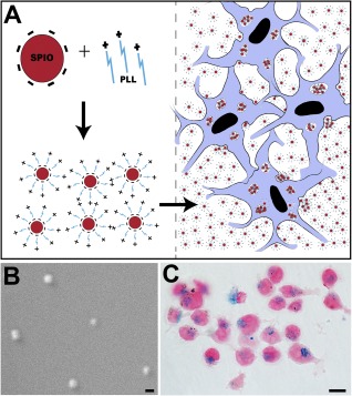
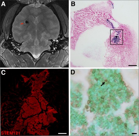
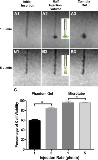
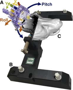
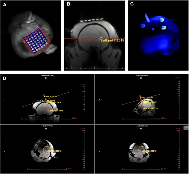
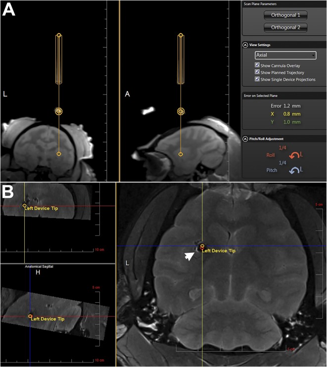
Optimal stem cell delivery procedures are critical to the success of the cell therapy approach. Variables such as flow rate, suspension solution, needle diameter, cell density, and tissue mechanics affect tissue penetration, backflow along the needle, and the dispersion and survival of injected cells during delivery. Most cell transplantation centers engaged in human clinical trials use custom-designed cannula needles, syringes, or catheters, sometimes precluding the use of magnetic resonance imaging (MRI)-guided delivery to target tissue. As a result, stem cell therapies may be hampered because more than 80% of grafted cells do not survive the delivery-for example, to the heart, liver/pancreas, and brain-which translates to poor patient outcomes. We developed a minimally invasive interventional MRI (iMRI) approach for intraoperatively imaging neural stem cell (NSC) delivery procedures. We used NSCs prelabeled with a contrast agent and real-time magnetic resonance imaging to guide the injection cannula to the target and to track the delivery of the cells into the putamen of baboons. We provide evidence that cell injection into the brain parenchyma follows a novel pulsatile mode of cellular discharge from the delivery catheter despite a constant infusion flow rate. The rate of cell infusion significantly affects the dispersion and viability of grafted cells. We report on our investigational use of a frameless navigation system for image-guided NSC transplantation using a straight cannula. Through submillimeter accuracy and real-time imaging, iMRI approaches may improve the safety and efficacy of neural cell transplantation therapies. Stem Cells Translational Medicine 2017;6:877-885.
优化的干细胞输送程序对细胞疗法的成功至关重要。流速、悬浮溶液、针头直径、细胞密度和组织力学等变量会影响组织穿透、针头沿程回流以及注射细胞在输送过程中的分散和存活。大多数从事人类临床试验的细胞移植中心使用定制设计的套管针、注射器或导管,有时会排除使用磁共振成像 (MRI) 引导输送到目标组织。结果,干细胞疗法可能会受到阻碍,因为超过 80%的移植细胞在输送过程中无法存活,例如输送到心脏、肝脏/胰腺和大脑,这导致患者的预后较差。我们开发了一种微创介入 MRI(iMRI)方法,用于术中成像神经干细胞(NSC)输送程序。我们使用预先用造影剂标记的 NSCs,并实时磁共振成像来引导注射套管针到达目标,并跟踪细胞输送到狒狒的壳核。我们提供的证据表明,尽管存在恒定的输注流速,但细胞注入脑实质遵循从输送导管中细胞释放的新型脉冲模式。细胞输注率显著影响移植细胞的分散和活力。我们报告了我们使用无框架导航系统进行直套管图像引导 NSC 移植的研究情况。通过亚毫米级精度和实时成像,iMRI 方法可能会提高神经细胞移植疗法的安全性和有效性。《干细胞转化医学》2017;6:877-885.