Department of Ophthalmology and Visual Sciences, University of Illinois at Chicago, College of Medicine, Chicago, IL, United States of America.
PLoS One. 2019 Jun 27;14(6):e0218879. doi: 10.1371/journal.pone.0218879. eCollection 2019.
We have previously reported that lamellar dissection of the cornea transects stromal nerves, and that regenerating neurites form a dense net along the surgical plane. In these experiments, we have disrupted the stromal nerve trunks in situ, without incising the cornea, to determine the regeneration events in the absence of a surgical plane.
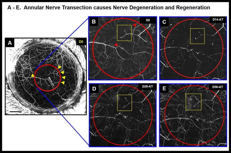
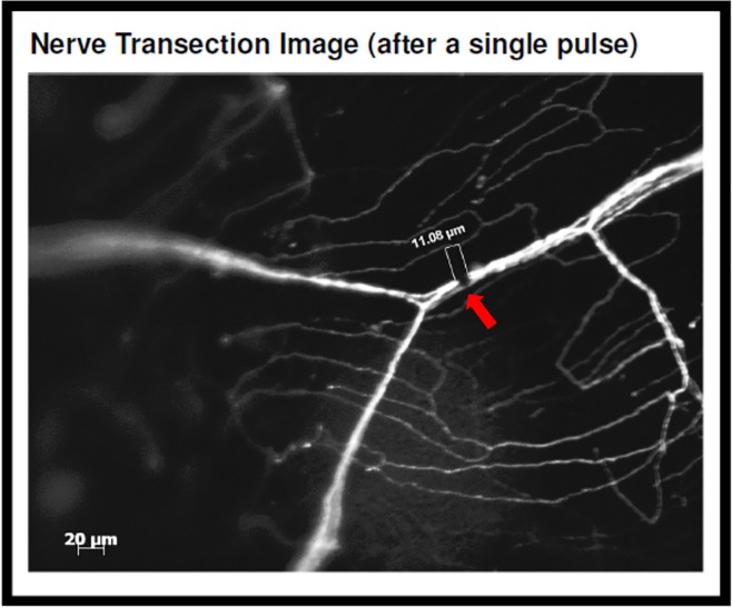
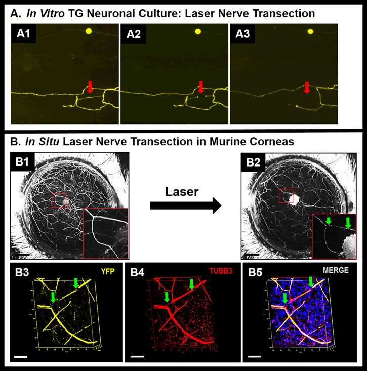
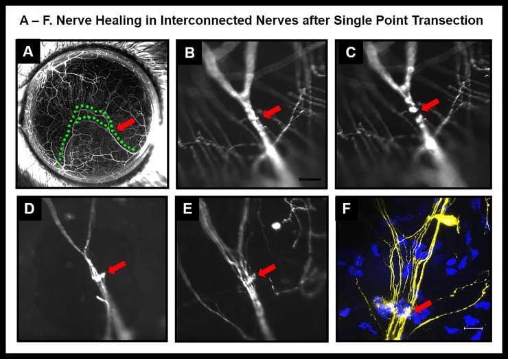
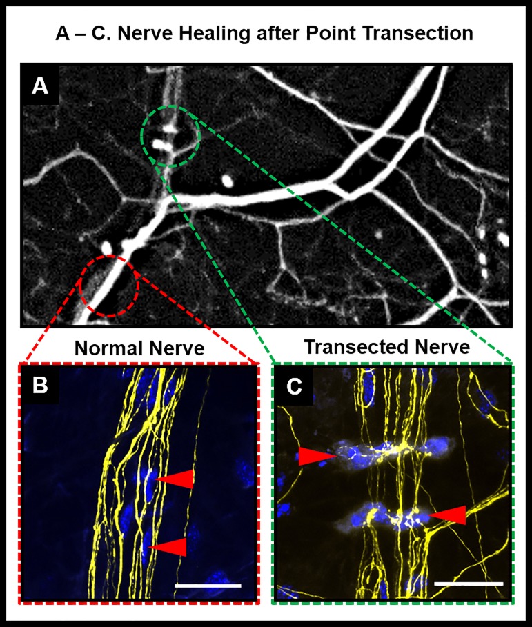
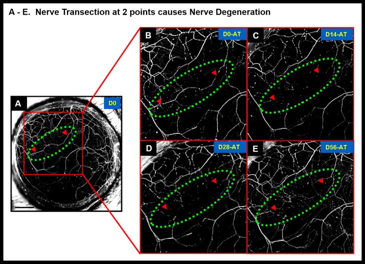
Thy1-YFP mice were anesthetized and in vivo images of the corneal nerves were obtained with a wide-field stereofluorescent microscope. A far infrared XYRCOS Laser attached to 20X objective of an upright microscope was used to perform in situ transection of the stromal nerves. 3 types of laser transections were performed (n = 5/group): (i) point transection (a single cut); (ii) segmental transection (two cuts enclosing a segment of nerve trunk); and (iii) annular transection (cuts on all nerve trunks crossing the perimeter of a 0.8 mm diameter circular area centered on the corneal apex). Mice were imaged sequentially for 4 weeks thereafter to assess nerve degeneration (disappearance or weakening of original fluorescence intensity) or regeneration (appearance of new fluorescent fronds). Beta-3-tubulin immunostaining was performed on corneal whole-mounts to demonstrate nerve disruption.
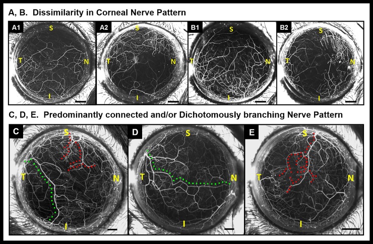
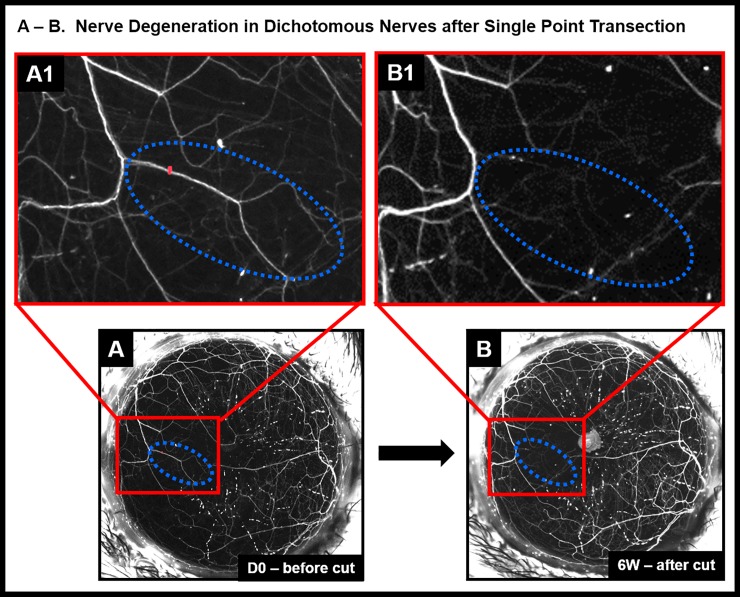
The pattern of stromal nerves in corneas of the same mouse and in corneas of littermates was dissimilar. Two distinct patterns were observed, often within the same cornea: (i) interconnected trunks that spanned limbus to limbus; or (ii) dichotomously branching trunks that terminate at the corneal apex. Point transections did not cause degeneration of proximal or distal segment in interconnected trunks, but resulted in degeneration of distal segment of branching trunks. In segmental transections, the nerve segment enclosed within the two laser cuts degenerated. Lack of beta-3 tubulin staining at transection site confirmed nerve transection. In interconnected trunks, at 4 weeks, a hyperfluorescent plaque filled the gap created by the transection. In annular transections, some nerve trunks degenerated, while others regained or retained fluorescence.
Interconnected stromal nerves in murine corneas do not degenerate after in situ point transection and show evidence of healing at the site of disruption. Presence or absence of a surgical plane influences corneal nerve regeneration after transection.
我们之前报道过角膜板层分离会切断基质神经,而且再生的神经突会沿着手术平面形成密集的网络。在这些实验中,我们在不切开角膜的情况下原位阻断基质神经干,以确定在没有手术平面的情况下的再生事件。
用宽场立体荧光显微镜对 Thy1-YFP 小鼠进行麻醉,并获得角膜神经的活体图像。远红外 XYRCOS 激光连接到直立显微镜的 20X 物镜上,用于进行基质神经的原位横切。进行了 3 种类型的激光横切(每组 n = 5):(i)点横切(单次切割);(ii)节段性横切(两次切割,包含一段神经干);和(iii)环形横切(在围绕角膜顶点的 0.8 毫米直径圆形区域的周边上的所有神经干上进行切割)。此后,对小鼠进行连续成像 4 周,以评估神经变性(原始荧光强度的消失或减弱)或再生(新荧光嫩枝的出现)。对角膜全层进行β-3-微管蛋白免疫染色以证明神经破坏。
同一只小鼠和同窝小鼠的角膜中基质神经的模式不同。通常在同一角膜中观察到两种不同的模式:(i)相互连接的干横跨角膜缘;或(ii)分叉分支的干终止于角膜顶点。点横切不会导致相互连接的干中的近端或远端节段变性,但会导致分支干的远端节段变性。在节段性横切中,被两个激光切割所包围的神经节段发生变性。横切部位缺乏β-3 微管蛋白染色证实了神经横切。在相互连接的干中,在 4 周时,一个高荧光斑块填充了横切所产生的间隙。在环形横切中,一些神经干变性,而其他神经干则恢复或保留荧光。
在原位点横切后,鼠角膜中的相互连接的基质神经不会变性,并显示出在破坏部位愈合的迹象。手术平面的存在与否会影响横切后角膜神经的再生。