Liu Chunyu, Tang Shenfei, Niu Guozhen, Zhang Juan, Huang Xinyu, Zhang Yushan, Bi Yanlong
Department of Ophthalmology, Tongji Hospital Affiliated with Tongji University School of Medicine, Shanghai 200333, P.R. China.
Exp Ther Med. 2019 Jul;18(1):242-252. doi: 10.3892/etm.2019.7573. Epub 2019 May 10.
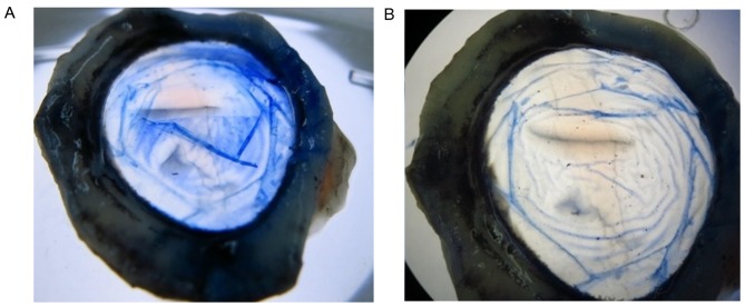
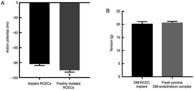
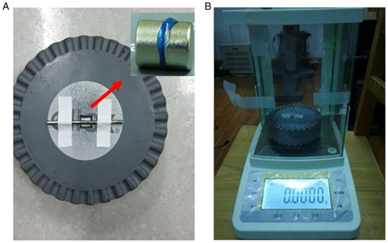
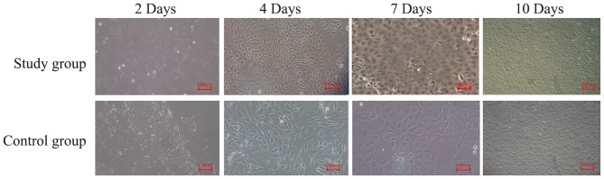
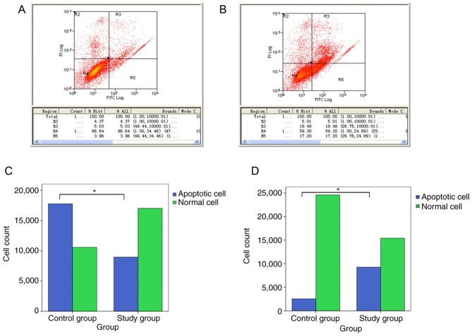
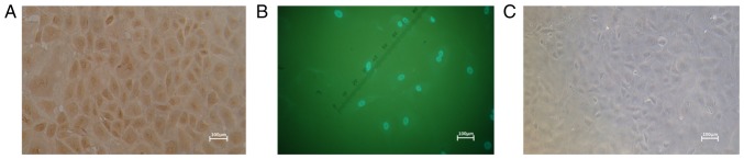
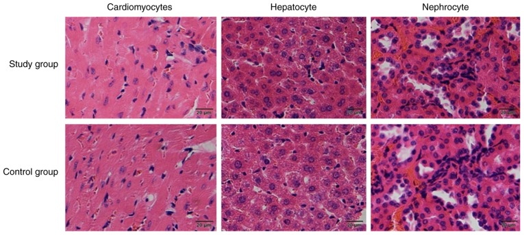
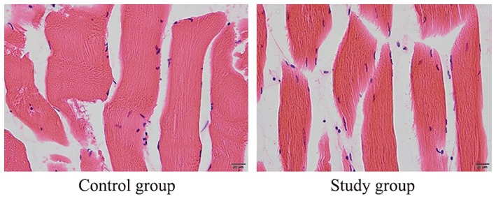
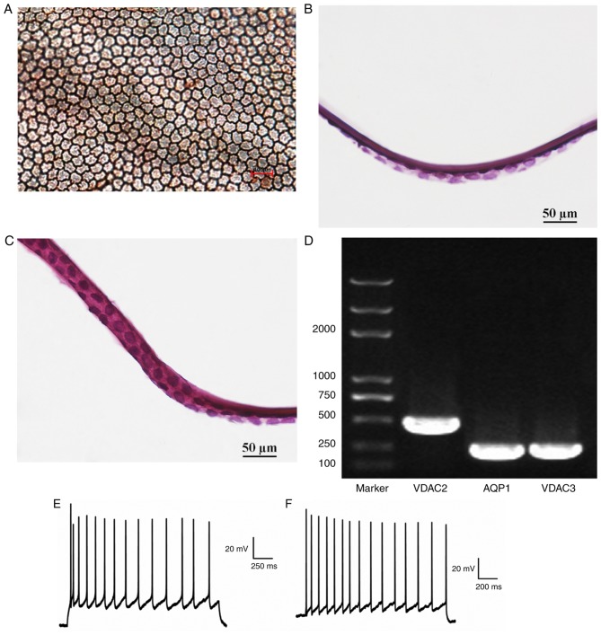
The aim of the present study was to investigate the feasibility of a new graft construction method using rabbit corneal endothelial cells (RCECs) and a porcine descemet membrane (DM) carrier. RCECs were isolated and the experimental group was treated with Y-27632, whereas the control group were cultured in medium without Y-27632. RCEC morphology was observed using an inverted microscope, and cell proliferation and apoptosis were detected by flow cytometry. To confirm the presence of RCECs, reverse transcription-quantitative PCR was used to detect gene expression levels of Na+-K+-ATPase, aquaporin 1, collagen α (IV), collagen α (VIII) and keratin-12. Histocompatibility testing was used to detect porcine DM antigenicity. A DM-RCEC graft was constructed, and morphology was observed using alizarin red-trypan blue and haematoxylin and eosin staining. Cell membrane potential was measured to evaluate the physical function of the DM-RCEC graft. Complex graft tension was measured using a modified tension detector and compared with fresh porcine DM-endothelium complex. -cultured RCECs formed a monolayer with a polygon morphology and cobblestone-like arrangement. -cultured RCECs exhibited typical RCEC characteristics before and after transplantation. The proliferation rates of the experimental and control groups were 62.68 and 34.50%, respectively (P<0.05); the apoptosis rates of the experimental and control groups were 8.99 and 35.68%, respectively (P<0.05). There was no antigenicity observed with the porcine DM. The action potential amplitude of the experimental and control groups was over -80 mV, reflecting normal RCEC physiological function. The tension measurements of the experimental and control groups were 20.0248±1.048 and 20.5013±0.657 g, respectively (P>0.05). Taken together, the results of the present study demonstrated that Y-27632 enhanced RCEC proliferation. In addition, the findings revealed the successful construction of a RCEC sheet on a porcine DM graft.
本研究的目的是探讨使用兔角膜内皮细胞(RCEC)和猪Descemet膜(DM)载体的新型移植片构建方法的可行性。分离RCEC,实验组用Y-27632处理,而对照组在不含Y-27632的培养基中培养。使用倒置显微镜观察RCEC形态,通过流式细胞术检测细胞增殖和凋亡。为了确认RCEC的存在,使用逆转录定量PCR检测Na+-K+-ATP酶、水通道蛋白1、胶原蛋白α(IV)、胶原蛋白α(VIII)和角蛋白-12的基因表达水平。使用组织相容性检测来检测猪DM的抗原性。构建DM-RCEC移植片,使用茜素红-台盼蓝和苏木精-伊红染色观察形态。测量细胞膜电位以评估DM-RCEC移植片的物理功能。使用改良的张力检测器测量复合移植片张力,并与新鲜猪DM-内皮复合体进行比较。Y-27632处理的RCEC形成具有多边形形态和鹅卵石样排列的单层。移植前后,Y-27632处理的RCEC表现出典型的RCEC特征。实验组和对照组的增殖率分别为62.68%和34.50%(P<0.05);实验组和对照组的凋亡率分别为8.99%和35.68%(P<0.05)。未观察到猪DM有抗原性。实验组和对照组的动作电位幅度超过-80 mV,反映RCEC生理功能正常。实验组和对照组的张力测量值分别为20.0248±1.048和20.5013±0.657 g(P>0.05)。综上所述,本研究结果表明Y-27632可促进RCEC增殖。此外,研究结果显示成功在猪DM移植片上构建了RCEC片层。