Musaeus Christian Sandøe, Nielsen Malene Schjønning, Høgh Peter
Department of Neurology, Danish Dementia Research Centre, Rigshospitalet, University of Copenhagen, Copenhagen, Denmark.
Regional Dementia Research Centre, Department of Neurology, Zealand University Hospital, Roskilde, Denmark.
Front Neurosci. 2019 Jun 11;13:563. doi: 10.3389/fnins.2019.00563. eCollection 2019.
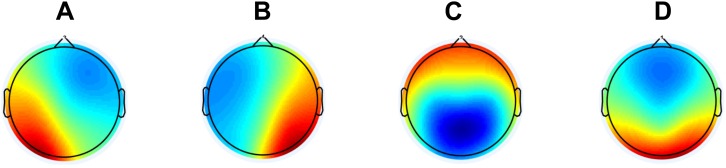
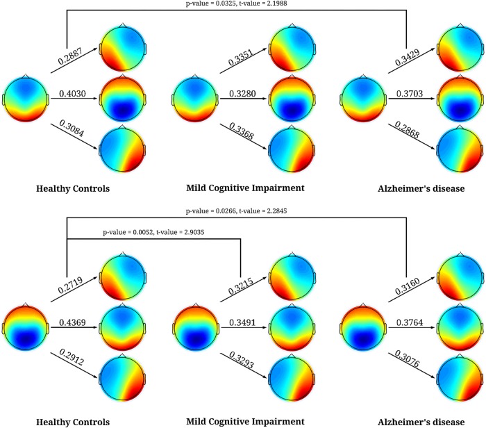
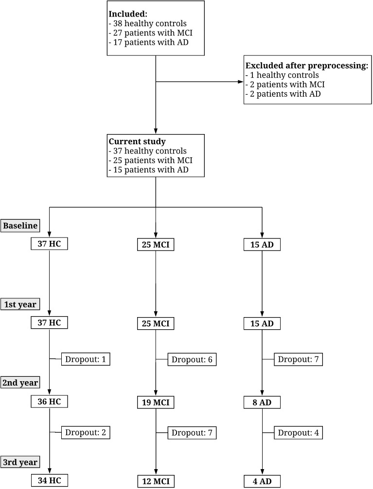
Network dysfunction is well established in patients with Alzheimer's disease (AD) and has been shown to be present early in the disease. This is especially interesting in patients with mild cognitive impairment (MCI) since they are more likely to develop AD. In EEG, one type of network analysis is microstates where the EEG is divided into quasi-stable states and these microstates have been linked to networks found with resting state functional MRI. In the current exploratory study, we therefore wanted to explore the changes in microstates in MCI, and AD compared to healthy controls (HC) and whether microstates were able to separate patients with MCI who progressed (pMCI) and those who remained stable (sMCI). EEGs were recorded at baseline for 17 patients with AD, 27 patients with MCI, and 38 older HC and the patients were followed for 3 years. To investigate whole-brain dynamics we extracted different microstate parameters. We found that patients with MCI, and AD had significantly higher occurrence (-value = 0.028), and coverage (-value = 0.010) for microstate A compared to HC. However, we did not find any significant systematic deviation of the transition probabilities from randomness for any of the groups. No significant differences were found between pMCI and sMCI but the largest difference in duration was found for microstate D. Microstate A has been linked to the temporal lobes in studies combining EEG and fMRI and the temporal lobes are the most affected by AD pathology in the early stages of the disease. This supports our idea that microstate A may be the first affected microstate in early AD. Even though not significant between pMCI and sMCI, Microstate D has previously been shown to be associated with both frontal and parietal areas as measured with fMRI and may correspond to underlying pathological changes in the progression of MCI to AD. However, larger studies are needed to confirm these findings.
网络功能障碍在阿尔茨海默病(AD)患者中已得到充分证实,并且已被证明在疾病早期就存在。这在轻度认知障碍(MCI)患者中尤其有趣,因为他们更有可能发展为AD。在脑电图(EEG)中,一种网络分析类型是微状态,其中EEG被分为准稳定状态,并且这些微状态已与静息态功能磁共振成像(fMRI)发现的网络相关联。因此,在当前的探索性研究中,我们想探讨MCI和AD患者与健康对照(HC)相比微状态的变化,以及微状态是否能够区分进展型MCI(pMCI)和稳定型MCI(sMCI)患者。在基线时记录了17例AD患者、27例MCI患者和38例老年HC的EEG,并对患者进行了3年的随访。为了研究全脑动力学,我们提取了不同的微状态参数。我们发现,与HC相比,MCI和AD患者的微状态A出现率(p值 = 0.028)和覆盖率(p值 = 0.010)显著更高。然而,我们没有发现任何一组的转移概率与随机性有任何显著的系统偏差。pMCI和sMCI之间没有发现显著差异,但微状态D的持续时间差异最大。在结合EEG和fMRI的研究中,微状态A与颞叶相关联,并且在疾病早期,颞叶是受AD病理影响最严重的部位。这支持了我们的观点,即微状态A可能是早期AD中第一个受影响的微状态。尽管pMCI和sMCI之间没有显著差异,但先前的研究表明,用fMRI测量时,微状态D与额叶和顶叶区域都有关联,并且可能对应于MCI进展为AD过程中的潜在病理变化。然而,需要更大规模的研究来证实这些发现。