• 文献检索
  • 文档翻译
  • 深度研究
  • 学术资讯
  • Suppr Zotero 插件Zotero 插件
  • 邀请有礼
  • 套餐&价格
  • 历史记录
应用&插件
Suppr Zotero 插件Zotero 插件浏览器插件Mac 客户端Windows 客户端微信小程序
定价
高级版会员购买积分包购买API积分包
服务
文献检索文档翻译深度研究API 文档MCP 服务
关于我们
关于 Suppr公司介绍联系我们用户协议隐私条款
关注我们

Suppr 超能文献

核心技术专利:CN118964589B侵权必究
粤ICP备2023148730 号-1Suppr @ 2026

文献检索

告别复杂PubMed语法,用中文像聊天一样搜索,搜遍4000万医学文献。AI智能推荐,让科研检索更轻松。

立即免费搜索

文件翻译

保留排版,准确专业,支持PDF/Word/PPT等文件格式,支持 12+语言互译。

免费翻译文档

深度研究

AI帮你快速写综述,25分钟生成高质量综述,智能提取关键信息,辅助科研写作。

立即免费体验

肺 F-FDG PET/HRCT 与基于计算机的 CT 分析协同应用于传统严重程度测量,以完善特发性肺纤维化(IPF)当前的风险分层。

Synergistic application of pulmonary F-FDG PET/HRCT and computer-based CT analysis with conventional severity measures to refine current risk stratification in idiopathic pulmonary fibrosis (IPF).

作者信息

Fraioli Francesco, Lyasheva Maria, Porter Joanna C, Bomanji Jamshed, Shortman Robert I, Endozo Raymond, Wan Simon, Bertoletti Linda, Machado Maria, Ganeshan Balaji, Win Thida, Groves Ashley M

机构信息

Institute of Nuclear Medicine, UCL(H) and University College London Hospital, 235 Euston Rd, London, NW1 2BU, UK.

Department of Oncology, School of Clinical Medicine, University of Cambridge, Cambridge, UK.

出版信息

Eur J Nucl Med Mol Imaging. 2019 Sep;46(10):2023-2031. doi: 10.1007/s00259-019-04386-5. Epub 2019 Jul 8.

DOI:10.1007/s00259-019-04386-5
PMID:31286201
原文链接:https://pmc.ncbi.nlm.nih.gov/articles/PMC6667685/
Abstract

INTRODUCTION

To investigate the combined performance of quantitative CT (qCT) following a computer algorithm analysis (IMBIO) and F-FDG PET/CT to assess survival in patients with idiopathic pulmonary fibrosis (IPF).

METHODS

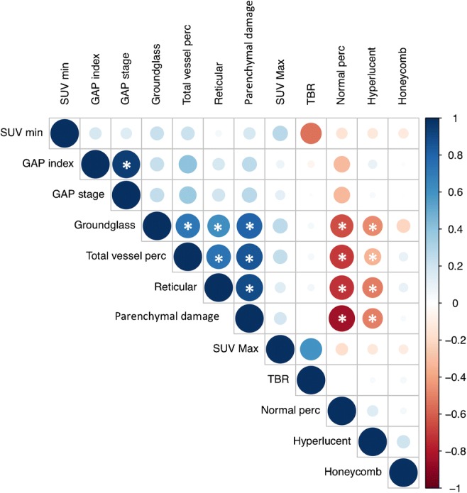
A total of 113 IPF patients (age 70 ± 9 years) prospectively and consecutively underwent F-FDG PET/CT and high-resolution CT (HRCT) at our institution. During a mean follow-up of 29.6 ± 26 months, 44 (48%) patients died. As part of the qCT analysis, pattern evaluation of HRCT (using IMBIO software) included the total extent (percentage) of the following features: normal-appearing lung, hyperlucent lung, parenchymal damage (comprising ground-glass opacification, reticular pattern and honeycombing), and the pulmonary vessels. The maximum (SUV) and minimum (SUV) standardized uptake value (SUV) for F-FDG uptake in the lungs, and the target-to-background (SUV/SUV) ratio (TBR) were quantified using routine region-of-interest (ROI) analysis. Pulmonary functional tests (PFTs) were acquired within 14 days of the PET/CT/HRCT scan. Kaplan-Meier (KM) survival analysis was used to identify associations with mortality.

RESULTS

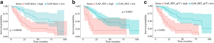
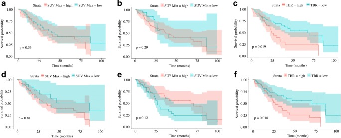
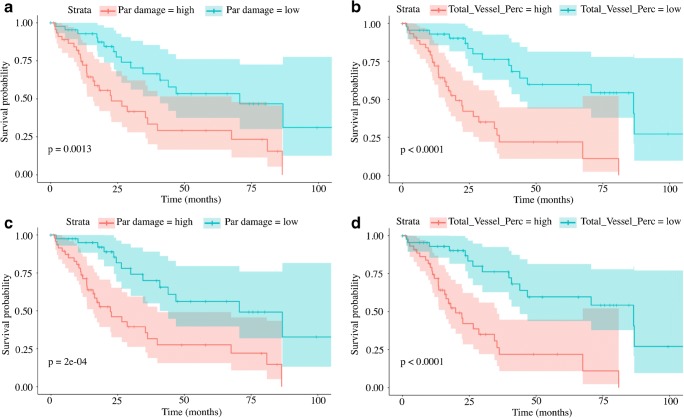
Data from 91 patients were available for comparative analysis. The average ± SD GAP [gender, age, physiology] score was 4.2 ± 1.7 (range 0-8). The average ± SD SUV, SUV, and TBR were 3.4 ± 1.4, 0.7 ± 0.2, and 5.6 ± 2.8, respectively. In all patients, qCT analysis demonstrated a predominantly reticular lung pattern (14.9 ± 12.4%). KM analysis showed that TBR (p = 0.018) and parenchymal damage assessed by qCT (p = 0.0002) were the best predictors of survival. Adding TBR and qCT to the GAP score significantly increased the ability to differentiate between high and low risk (p < 0.0001).

CONCLUSION

F-FDG PET and qCT are independent and synergistic in predicting mortality in patients with IPF.

摘要

引言

研究定量CT(qCT)结合计算机算法分析(IMBIO)和F-FDG PET/CT评估特发性肺纤维化(IPF)患者生存率的联合性能。

方法

共有113例IPF患者(年龄70±9岁)在我院前瞻性、连续地接受了F-FDG PET/CT和高分辨率CT(HRCT)检查。在平均29.6±26个月的随访期间,44例(48%)患者死亡。作为qCT分析的一部分,HRCT的模式评估(使用IMBIO软件)包括以下特征的总范围(百分比):外观正常的肺、肺透亮度增加、实质损害(包括磨玻璃影、网状影和蜂窝状影)以及肺血管。使用常规感兴趣区(ROI)分析对肺内F-FDG摄取的最大(SUV)和最小(SUV)标准化摄取值(SUV)以及靶本比(SUV/SUV)(TBR)进行量化。在PET/CT/HRCT扫描后14天内进行肺功能测试(PFT)。采用Kaplan-Meier(KM)生存分析来确定与死亡率的关联。

结果

91例患者的数据可用于比较分析。平均±标准差GAP[性别、年龄、生理学]评分为4.2±1.7(范围0-8)。平均±标准差SUV、SUV和TBR分别为3.4±1.4、0.7±0.2和5.6±2.8。在所有患者中,qCT分析显示主要为网状肺模式(14.9±12.4%)。KM分析表明,TBR(p=0.018)和qCT评估的实质损害(p=0.0002)是生存的最佳预测指标。将TBR和qCT添加到GAP评分中显著提高了区分高风险和低风险的能力(p<0.0001)。

结论

F-FDG PET和qCT在预测IPF患者死亡率方面具有独立性和协同性。

https://cdn.ncbi.nlm.nih.gov/pmc/blobs/88a4/6667685/8540384a5afe/259_2019_4386_Fig4_HTML.jpg
https://cdn.ncbi.nlm.nih.gov/pmc/blobs/88a4/6667685/66cd45730a66/259_2019_4386_Fig1_HTML.jpg
https://cdn.ncbi.nlm.nih.gov/pmc/blobs/88a4/6667685/513e7daec172/259_2019_4386_Fig2_HTML.jpg
https://cdn.ncbi.nlm.nih.gov/pmc/blobs/88a4/6667685/9f5fabc20a1b/259_2019_4386_Fig3_HTML.jpg
https://cdn.ncbi.nlm.nih.gov/pmc/blobs/88a4/6667685/8540384a5afe/259_2019_4386_Fig4_HTML.jpg

相似文献

1
Synergistic application of pulmonary F-FDG PET/HRCT and computer-based CT analysis with conventional severity measures to refine current risk stratification in idiopathic pulmonary fibrosis (IPF).肺 F-FDG PET/HRCT 与基于计算机的 CT 分析协同应用于传统严重程度测量,以完善特发性肺纤维化(IPF)当前的风险分层。
Eur J Nucl Med Mol Imaging. 2019 Sep;46(10):2023-2031. doi: 10.1007/s00259-019-04386-5. Epub 2019 Jul 8.
2
Pulmonary F-FDG uptake helps refine current risk stratification in idiopathic pulmonary fibrosis (IPF).肺 F-FDG 摄取有助于细化特发性肺纤维化 (IPF) 中的当前风险分层。
Eur J Nucl Med Mol Imaging. 2018 May;45(5):806-815. doi: 10.1007/s00259-017-3917-8. Epub 2018 Jan 16.
3
Idiopathic pulmonary fibrosis and diffuse parenchymal lung disease: implications from initial experience with 18F-FDG PET/CT.特发性肺纤维化和弥漫性实质性肺疾病:18F-FDG PET/CT 初步经验的启示
J Nucl Med. 2009 Apr;50(4):538-45. doi: 10.2967/jnumed.108.057901. Epub 2009 Mar 16.
4
[F]FDG PET/CT predicts progression-free survival in patients with idiopathic pulmonary fibrosis.[F]氟代脱氧葡萄糖正电子发射断层扫描/计算机断层扫描(FDG PET/CT)可预测特发性肺纤维化患者的无进展生存期。
Respir Res. 2017 Apr 27;18(1):74. doi: 10.1186/s12931-017-0556-3.
5
The Value of F-FDG PET/CT in Evaluating Disease Severity and Prognosis in Idiopathic Pulmonary Fibrosis Patients.F-FDG PET/CT 在评估特发性肺纤维化患者疾病严重程度和预后中的价值。
J Korean Med Sci. 2021 Oct 25;36(41):e257. doi: 10.3346/jkms.2021.36.e257.
6
FDG-PET/CT in the prediction of pulmonary function improvement in nonspecific interstitial pneumonia. A Pilot Study.氟代脱氧葡萄糖正电子发射断层扫描/计算机断层扫描在预测非特异性间质性肺炎肺功能改善中的应用。一项初步研究。
Eur J Radiol. 2016 Dec;85(12):2200-2205. doi: 10.1016/j.ejrad.2016.10.001. Epub 2016 Oct 4.
7
Imaging Pulmonary Fibrosis and Treatment Efficacy In Vivo with Autotaxin-Specific PET Ligand [F]ATX-1905.用自主分泌酶特异性 PET 配体 [F]ATX-1905 进行肺纤维化的体内成像和治疗效果评估。
Mol Pharm. 2024 Oct 7;21(10):5171-5181. doi: 10.1021/acs.molpharmaceut.4c00571. Epub 2024 Aug 26.
8
Can FDG-PET/CT imaging be used to predict decline in quality of life in interstitial lung disease? A prospective study of the relationship between FDG uptake and quality of life in a UK outpatient setting.FDG-PET/CT 成像能否用于预测间质性肺疾病患者生活质量下降?一项英国门诊环境中 FDG 摄取与生活质量关系的前瞻性研究。
BMJ Open. 2024 May 30;14(5):e081103. doi: 10.1136/bmjopen-2023-081103.
9
More advantages in detecting bone and soft tissue metastases from prostate cancer using F-PSMA PET/CT.使用F-PSMA PET/CT检测前列腺癌骨和软组织转移方面有更多优势。
Hell J Nucl Med. 2019 Jan-Apr;22(1):6-9. doi: 10.1967/s002449910952. Epub 2019 Mar 7.
10
Combination of positron emission tomography/computed tomography and chest thin-layer high-resolution computed tomography for evaluation of pulmonary nodules: Correlation with imaging features, maximum standardized uptake value, and pathology.正电子发射断层扫描/计算机断层扫描与胸部薄层高分辨率计算机断层扫描联合用于评估肺结节:与影像学特征、最大标准化摄取值及病理的相关性
Medicine (Baltimore). 2018 Aug;97(31):e11640. doi: 10.1097/MD.0000000000011640.

引用本文的文献

1
Ga-FAPI PET/CT for diagnostic accuracy and therapeutic response assessment in bleomycin-induced pulmonary fibrosis: an integrated preclinical study.镓标记的成纤维细胞活化蛋白抑制剂正电子发射断层扫描/计算机断层扫描在博来霉素诱导的肺纤维化诊断准确性及治疗反应评估中的应用:一项综合性临床前研究
Front Med (Lausanne). 2025 Jun 26;12:1613010. doi: 10.3389/fmed.2025.1613010. eCollection 2025.
2
Targeted imaging of pulmonary fibrosis by a cyclic peptide LyP-1.环肽LyP-1对肺纤维化的靶向成像
Sci Rep. 2025 Mar 8;15(1):8098. doi: 10.1038/s41598-024-78068-0.
3
Molecular Imaging of Pulmonary Fibrosis.

本文引用的文献

1
Pulmonary F-FDG uptake helps refine current risk stratification in idiopathic pulmonary fibrosis (IPF).肺 F-FDG 摄取有助于细化特发性肺纤维化 (IPF) 中的当前风险分层。
Eur J Nucl Med Mol Imaging. 2018 May;45(5):806-815. doi: 10.1007/s00259-017-3917-8. Epub 2018 Jan 16.
2
Clinical trial research in focus: why do so many clinical trials fail in IPF?聚焦临床试验研究:为何众多特发性肺纤维化临床试验会失败?
Lancet Respir Med. 2017 May;5(5):372-374. doi: 10.1016/S2213-2600(17)30122-4. Epub 2017 Apr 12.
3
Impact of pulmonary vascular volume on mortality in IPF: is it time to reconsider the role of vasculature in disease pathogenesis and progression?
肺纤维化的分子成像
J Nucl Med. 2025 Apr 1;66(4):502-505. doi: 10.2967/jnumed.124.267852.
4
[F]AlF-CBP imaging of type I collagen for non-invasive monitoring of pulmonary fibrosis in preclinical models.利用 AlF-CBP 对 I 型胶原进行成像,以实现对临床前模型中肺纤维化的非侵入性监测。
Eur J Nucl Med Mol Imaging. 2024 Dec;52(1):22-35. doi: 10.1007/s00259-024-06888-3. Epub 2024 Aug 22.
5
[Ga]FAPI PET for Imaging and Treatment Monitoring in a Preclinical Model of Pulmonary Fibrosis: Comparison to [F]FDG PET and CT.[镓]FAPI PET用于肺纤维化临床前模型的成像和治疗监测:与[氟]FDG PET及CT的比较
Pharmaceuticals (Basel). 2024 Jun 3;17(6):726. doi: 10.3390/ph17060726.
6
Imaging at the nexus: how state of the art imaging techniques can enhance our understanding of cancer and fibrosis.影像学在其中的作用:最先进的影像学技术如何增强我们对癌症和纤维化的理解。
J Transl Med. 2024 Jun 13;22(1):567. doi: 10.1186/s12967-024-05379-1.
7
[F]FDG PET/CT Signal Correlates with Neoangiogenesis Markers in Patients with Fibrotic Interstitial Lung Disease Who Underwent Lung Biopsy: Implication for the Use of PET/CT in Diffuse Lung Diseases.[F]FDG PET/CT 信号与接受肺活检的纤维化间质性肺病患者的新生血管生成标志物相关:对弥漫性肺疾病中使用 PET/CT 的影响。
J Nucl Med. 2024 Apr 1;65(4):617-622. doi: 10.2967/jnumed.123.266445.
8
Assessing robustness of quantitative susceptibility-based MRI radiomic features in patients with multiple sclerosis.评估多发性硬化症患者基于定量磁化率磁共振成像(QS-MRI)放射组学特征的稳健性。
Sci Rep. 2023 Sep 27;13(1):16239. doi: 10.1038/s41598-023-42914-4.
9
Acute Exacerbations of Interstitial Lung Diseases: Focus on Biomarkers.间质性肺疾病的急性加重:聚焦生物标志物。
Int J Mol Sci. 2023 Jun 15;24(12):10196. doi: 10.3390/ijms241210196.
10
Radiolabeled GPVI-Fc for PET Imaging of Multiple Extracellular Matrix Fibers: A New Look into Pulmonary Fibrosis Progression.用于多种细胞外基质纤维PET成像的放射性标记GPVI-Fc:对肺纤维化进展的新认识。
J Nucl Med. 2023 Jun;64(6):940-945. doi: 10.2967/jnumed.122.264552. Epub 2023 Jan 26.
肺血管容量对特发性肺纤维化死亡率的影响:是否是时候重新审视血管系统在疾病发病机制和进展中的作用了?
Eur Respir J. 2017 Feb 23;49(2). doi: 10.1183/13993003.02345-2016. Print 2017 Feb.
4
Quantification of Lung PET Images: Challenges and Opportunities.肺部PET图像的定量分析:挑战与机遇
J Nucl Med. 2017 Feb;58(2):201-207. doi: 10.2967/jnumed.116.184796. Epub 2017 Jan 12.
5
Automated Quantitative Computed Tomography Versus Visual Computed Tomography Scoring in Idiopathic Pulmonary Fibrosis: Validation Against Pulmonary Function.特发性肺纤维化中自动定量计算机断层扫描与视觉计算机断层扫描评分的比较:与肺功能的对照验证
J Thorac Imaging. 2016 Sep;31(5):304-11. doi: 10.1097/RTI.0000000000000220.
6
Improved correction for the tissue fraction effect in lung PET/CT imaging.改善肺部PET/CT成像中组织分数效应的校正。
Phys Med Biol. 2015 Sep 21;60(18):7387-402. doi: 10.1088/0031-9155/60/18/7387. Epub 2015 Sep 9.
7
Areas of normal pulmonary parenchyma on HRCT exhibit increased FDG PET signal in IPF patients.特发性肺纤维化患者 HRCT 正常肺实质区域的 FDG PET 信号增加。
Eur J Nucl Med Mol Imaging. 2014 Feb;41(2):337-42. doi: 10.1007/s00259-013-2514-8. Epub 2013 Aug 14.
8
Interobserver variability in the CT assessment of honeycombing in the lungs.肺部蜂窝状改变的 CT 评估中的观察者间变异性。
Radiology. 2013 Mar;266(3):936-44. doi: 10.1148/radiol.12112516. Epub 2012 Dec 6.
9
Idiopathic pulmonary fibrosis and diffuse parenchymal lung disease: implications from initial experience with 18F-FDG PET/CT.特发性肺纤维化和弥漫性实质性肺疾病:18F-FDG PET/CT 初步经验的启示
J Nucl Med. 2009 Apr;50(4):538-45. doi: 10.2967/jnumed.108.057901. Epub 2009 Mar 16.
10
Imaging pulmonary inflammation with positron emission tomography: a biomarker for drug development.利用正电子发射断层扫描成像肺部炎症:药物研发的生物标志物
Mol Pharm. 2006 Sep-Oct;3(5):488-95. doi: 10.1021/mp060050w.