Miccoli Beatrice, Lopez Carolina Mora, Goikoetxea Erkuden, Putzeys Jan, Sekeri Makrina, Krylychkina Olga, Chang Shuo-Wen, Firrincieli Andrea, Andrei Alexandru, Reumers Veerle, Braeken Dries
IMEC, Heverlee, Belgium.
KU Leuven, Leuven, Belgium.
Front Neurosci. 2019 Jun 25;13:641. doi: 10.3389/fnins.2019.00641. eCollection 2019.
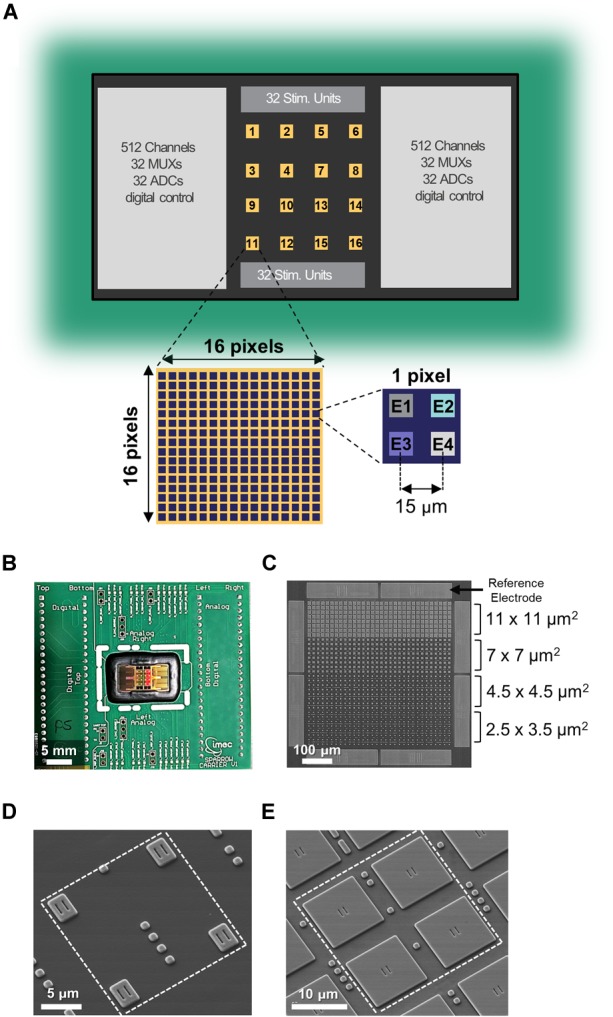
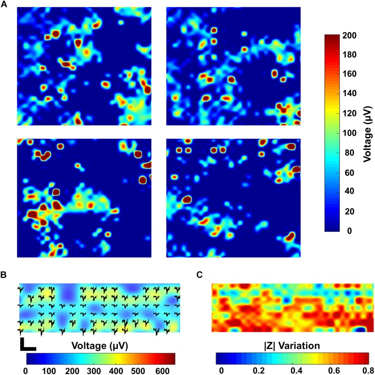
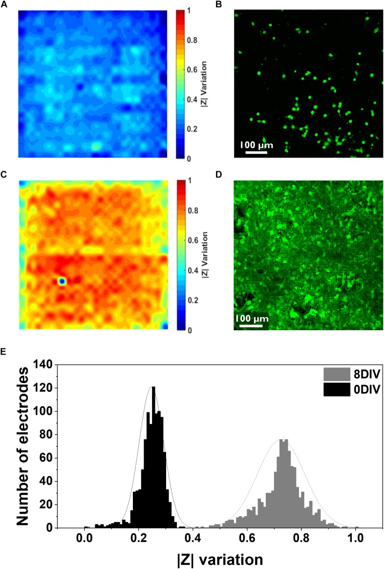
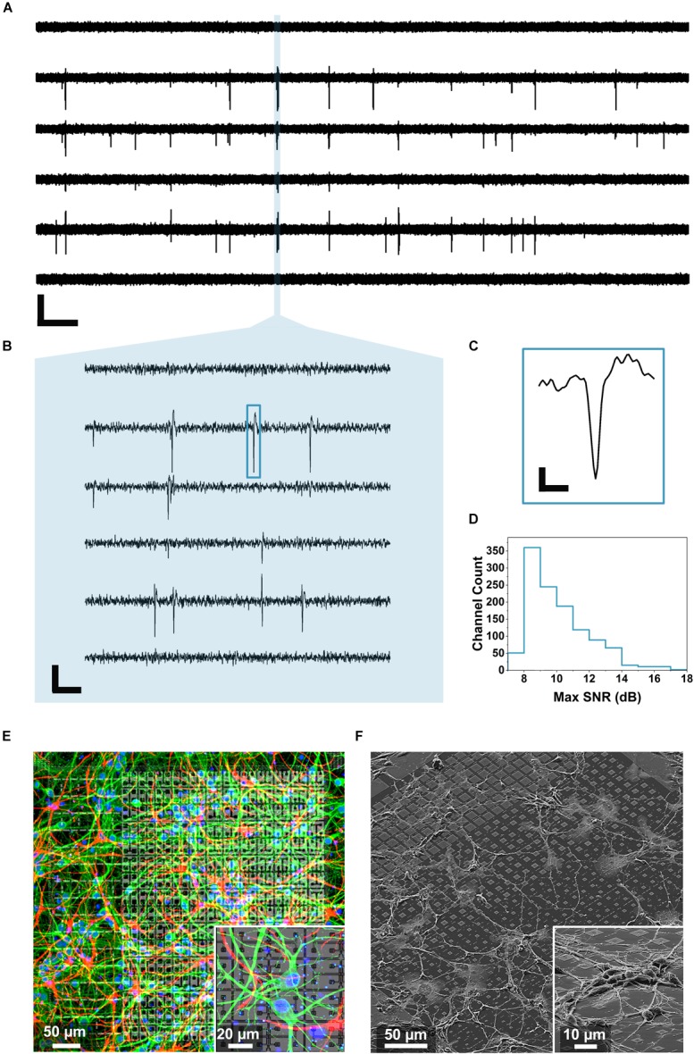
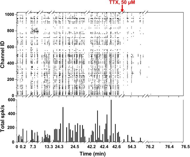
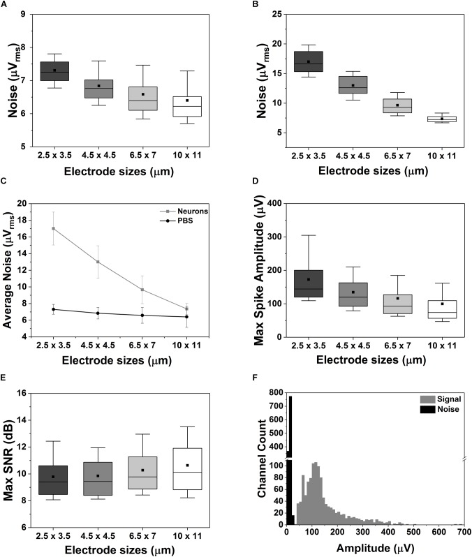
Multi-electrode arrays, both active or passive, emerged as ideal technologies to unveil intricated electrophysiological dynamics of cells and tissues. Active MEAs, designed using complementary metal oxide semiconductor technology (CMOS), stand over passive devices thanks to the possibility of achieving single-cell resolution, the reduced electrode size, the reduced crosstalk and the higher functionality and portability. Nevertheless, most of the reported CMOS MEA systems mainly rely on a single operational modality, which strongly hampers the applicability range of a single device. This can be a limiting factor considering that most biological and electrophysiological dynamics are often based on the synergy of multiple and complex mechanisms acting from different angles on the same phenomena. Here, we designed a CMOS MEA chip with 16,384 titanium nitride electrodes, 6 independent operational modalities and 1,024 parallel recording channels for neuro-electrophysiological studies. Sixteen independent active areas are patterned on the chip surface forming a 4 × 4 matrix, each one including 1,024 electrodes. Electrodes of four different sizes are present on the chip surface, ranging from 2.5 × 3.5 μm up to 11 × 11.0 μm, with 15 μm pitch. In this paper, we exploited the impedance monitoring and voltage recording modalities not only to monitor the growth and development of primary rat hippocampal neurons, but also to assess their electrophysiological activity over time showing a mean spike amplitude of 144.8 ± 84.6 μV. Fixed frequency (1 kHz) and high sampling rate (30 kHz) impedance measurements were used to evaluate the cellular adhesion and growth on the chip surface. Thanks to the high-density configuration of the electrodes, as well as their dimension and pitch, the chip can appreciate the evolutions of the cell culture morphology starting from the moment of the seeding up to mature culture conditions. The measurements were confirmed by fluorescent staining. The effect of the different electrode sizes on the spike amplitudes and noise were also discussed. The multi-modality of the presented CMOS MEA allows for the simultaneous assessment of different physiological properties of the cultured neurons. Therefore, it can pave the way both to answer complex fundamental neuroscience questions as well as to aid the current drug-development paradigm.
多电极阵列,无论是有源的还是无源的,都已成为揭示细胞和组织复杂电生理动力学的理想技术。采用互补金属氧化物半导体技术(CMOS)设计的有源微电极阵列(MEA)优于无源器件,因为它能够实现单细胞分辨率、减小电极尺寸、减少串扰以及具有更高的功能性和便携性。然而,大多数已报道的CMOS MEA系统主要依赖单一的操作模式,这严重限制了单个设备的适用范围。考虑到大多数生物和电生理动力学通常基于多种复杂机制在同一现象上从不同角度的协同作用,这可能是一个限制因素。在此,我们设计了一种用于神经电生理研究的CMOS MEA芯片,该芯片具有16384个氮化钛电极、6种独立的操作模式和1024个并行记录通道。16个独立的有源区域在芯片表面形成一个4×4矩阵,每个区域包含1024个电极。芯片表面存在四种不同尺寸的电极,范围从2.5×3.5μm到11×11.0μm,间距为15μm。在本文中,我们不仅利用阻抗监测和电压记录模式来监测原代大鼠海马神经元的生长和发育,还评估了它们随时间的电生理活动,平均峰电位幅度为144.8±84.6μV。使用固定频率(1kHz)和高采样率(30kHz)的阻抗测量来评估细胞在芯片表面的粘附和生长。由于电极的高密度配置以及它们的尺寸和间距,该芯片能够从接种时刻到成熟培养条件下观察细胞培养形态的演变。测量结果通过荧光染色得到证实。还讨论了不同电极尺寸对峰电位幅度和噪声的影响。所展示的CMOS MEA的多模式特性允许同时评估培养神经元的不同生理特性。因此,它既可以为回答复杂的基础神经科学问题铺平道路,也有助于当前的药物开发模式。