Li Yuanzhi, Guo Shenquan, Liu Wenchao, Jin Tao, Li Xifeng, He Xuying, Zhang Xin, Su Hengxian, Zhang Nan, Duan Chuanzhi
The National Key Clinical Specialty, The Engineering Technology Research Center of Education Ministry of China, Guangdong Provincial Key Laboratory on Brain Function Repair and Regeneration, Department of Neurosurgery, Zhujiang Hospital, Southern Medical University, Guangzhou, China.
Department of Neurosurgery, Affiliated Hengyang Hospital, Southern Medical University (Hengyang Central Hospital), Hengyang, China.
Front Neurosci. 2019 Jun 25;13:645. doi: 10.3389/fnins.2019.00645. eCollection 2019.
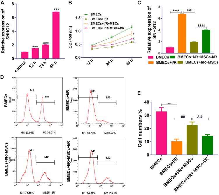
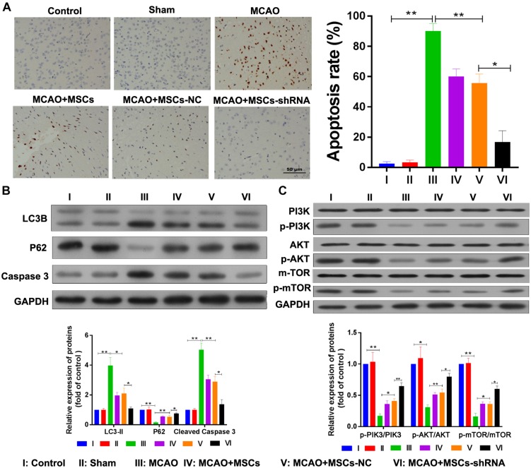
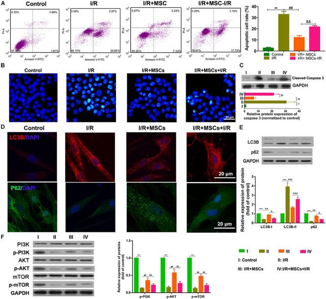
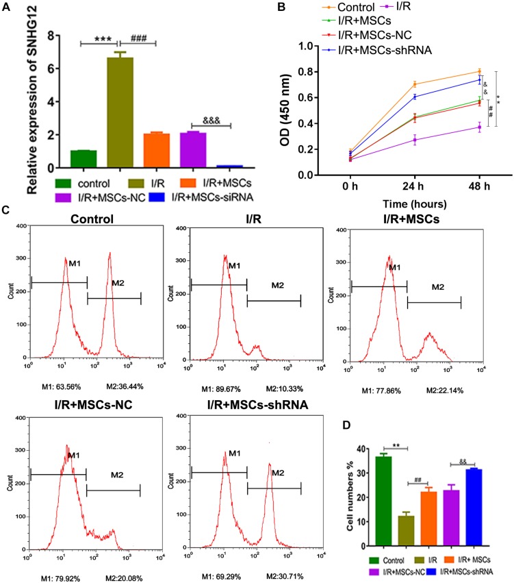
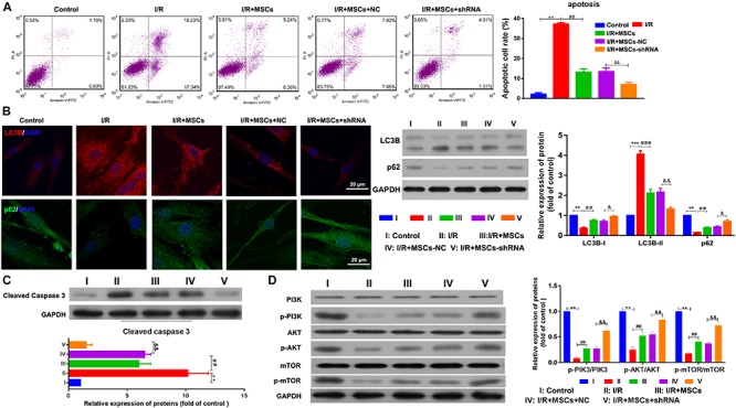
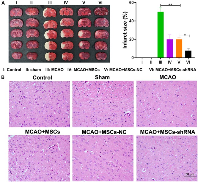
Previous studies have reported that the long non-coding RNA SNHG12 (lncRNA SNHG12) plays a critical role in regulating the function of mesenchymal stem cells (MSCs); however, the effect of lncRNA SNHG12 on MSCs in injured brain tissue has rarely been reported. We studied the effect and mechanism of lncRNA SNHG12-modified mesenchymal stem cells (MSCs) in treating brain injuries caused by ischemia/reperfusion (I/R). I/R treated rat brain microvascular endothelial cells (BMECs) were co-cultured with MSCs or I/R pretreated MSCs. Next, BMEC proliferation was detected by using CCK-8 and EdU assays, and cell apoptosis was determined by using flow cytometry and the Hoechst staining method. Autophagy of BMECs was determined using immunofluorescence and expression of associated pathway proteins were measured by western blotting. Moreover, BMEC proliferation, apoptosis, and autophagy were also determined after the BMECs had been co-cultured with shSNHG12-MSCs. In addition, a rat model of middle cerebral artery occlusion (MCAO) was used to further confirm the findings obtained with cells. I/R treatment significantly decreased the proliferation of BMECs, but increased their levels of SNHG12 expression, apoptosis, and autophagy. However, co-culturing of BMECs with MSCs markedly alleviated the reduction in BMEC proliferation and the increases in BMEC apoptosis and autophagy, as well as the phosphorylation of PI3K, AKT, and mTOR proteins in BMECs that had been induced by I/R. Furthermore, shSNHG12 remarkably enhanced the effects of MSCs. In addition, an injection MSCs reduced the infarct areas and rates of cell apoptosis in MACO rats, and reduced the phosphorylation of PI3K, AKT, and mTOR proteins. Moreover, shSNHG12 enhanced the ameliorative effect of MSCs in treating brain injuries in the MACO rats. In conclusion, silencing of SNHG12 enhanced the effects of MSCs in reducing apoptosis and autophagy of BMECs by activating the PI3K/AKT/mTOR signaling pathway.
以往研究报道,长链非编码RNA SNHG12(lncRNA SNHG12)在调节间充质干细胞(MSC)功能中起关键作用;然而,lncRNA SNHG12对损伤脑组织中MSC的影响鲜有报道。我们研究了lncRNA SNHG12修饰的间充质干细胞(MSC)在治疗缺血/再灌注(I/R)所致脑损伤中的作用及机制。将I/R处理的大鼠脑微血管内皮细胞(BMEC)与MSC或I/R预处理的MSC共培养。接下来,使用CCK-8和EdU检测法检测BMEC增殖,使用流式细胞术和Hoechst染色法测定细胞凋亡。使用免疫荧光法测定BMEC的自噬,并通过蛋白质印迹法测量相关信号通路蛋白的表达。此外,将BMEC与shSNHG12-MSC共培养后,也测定了BMEC的增殖、凋亡和自噬。此外,使用大脑中动脉闭塞(MCAO)大鼠模型进一步证实细胞实验结果。I/R处理显著降低了BMEC的增殖,但增加了其SNHG12表达水平、凋亡和自噬。然而,BMEC与MSC共培养显著减轻了I/R诱导的BMEC增殖减少、BMEC凋亡和自噬增加,以及BMEC中PI3K、AKT和mTOR蛋白的磷酸化。此外,shSNHG12显著增强了MSC的作用。此外,注射MSC可减少MCAO大鼠的梗死面积和细胞凋亡率,并降低PI3K、AKT和mTOR蛋白的磷酸化。此外,shSNHG12增强了MSC对MCAO大鼠脑损伤的改善作用。总之,沉默SNHG12可通过激活PI3K/AKT/mTOR信号通路增强MSC减少BMEC凋亡和自噬的作用。