Biomechanics Laboratory, Department of Electrical and Mechanical Engineering, Graduate School of Engineering, Nagoya Institute of Technology, Gokiso-cho, Showa-ku, Nagoya, 466-8555, Japan.
Biomech Model Mechanobiol. 2020 Feb;19(1):147-157. doi: 10.1007/s10237-019-01201-w. Epub 2019 Jul 11.
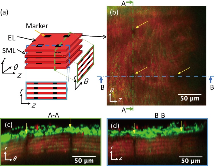
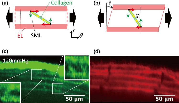
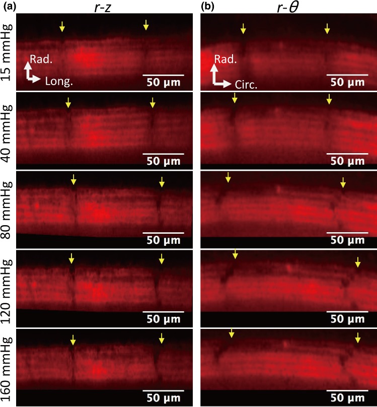
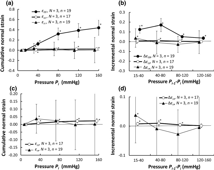
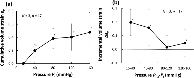
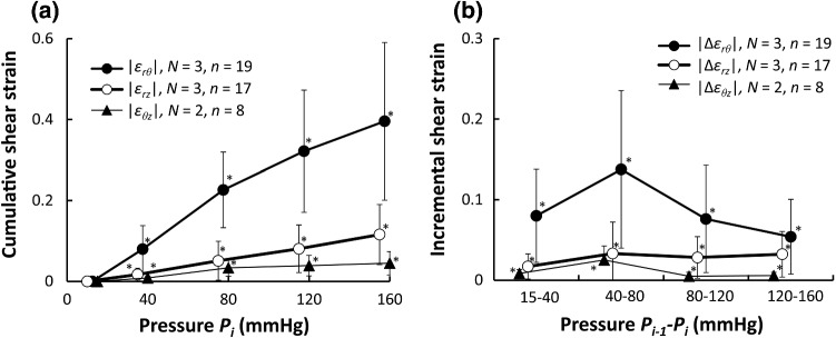
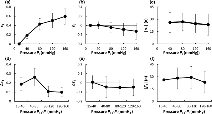
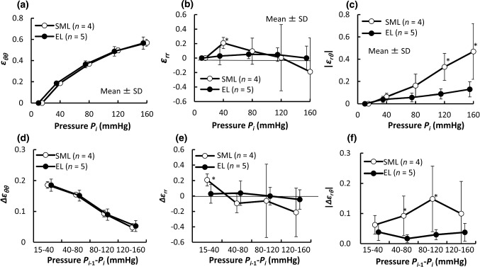
The aorta is composed of various constituents with different mechanical properties. This heterogeneous structure implies non-uniform deformation in the aorta, which could affect local cell functions. The present study investigates 3D strains of the aorta at a cell scale induced by intraluminal pressurization. After resected mouse, thoracic aortas were stretched to their in vivo length, and the aortas were pressurized at 15, 40, 80, 120, and 160 mmHg. Images of autofluorescent light of elastin were captured under a two-photon microscope. From the movement of markers in elastic laminas (ELs) created by photo-bleaching, 3D strains (ε, ε, ε, ε, ε, ε) between two neighboring ELs in the circumferential (θ), longitudinal (z), and radial (r) directions with reference to the dimensions at 15 mmHg were calculated. The results demonstrated that the average of shear strain ε was almost 0 in a physiological pressure range (from 80 to 120 mmHg) with an absolute value |ε| changing approximately by 5%. This indicates that ELs experience radial-circumferential shear at the cell scale, but not at the whole tissue scale. The normal strains in the circumferential ε and longitudinal direction ε were positive but that in the radial direction ε was almost 0, which demonstrates that aortic tissue is not an incompressible material. The first principal direction in the radial-circumferential plane was 29° ± 13° from the circumferential direction. We show that the aorta is not simply stretched in the circumferential direction during pressurization and that cells in the aorta undergo complex deformations by nature.
主动脉由具有不同机械性能的各种成分组成。这种不均匀的结构意味着主动脉的变形不均匀,这可能会影响局部细胞功能。本研究研究了腔内加压引起的主动脉在细胞尺度上的三维应变。在切除小鼠后,将胸主动脉拉伸至其体内长度,并在 15、40、80、120 和 160mmHg 下对主动脉加压。在双光子显微镜下拍摄弹性蛋白自发荧光的图像。通过光漂白产生的弹性层(EL)中的标记的运动,计算了在圆周(θ)、纵向(z)和径向(r)方向上两个相邻 EL 之间的三维应变(ε,ε,ε,ε,ε,ε)相对于 15mmHg 时的尺寸。结果表明,在生理压力范围内(80 至 120mmHg),剪切应变ε的平均值几乎为 0,绝对值|ε|的变化约为 5%。这表明 EL 在细胞尺度上经历径向-周向剪切,但不在整个组织尺度上。周向应变ε和纵向应变ε为正,但径向应变ε几乎为 0,这表明主动脉组织不是不可压缩的材料。径向-周向平面中的第一主方向与周向方向相差 29°±13°。我们表明,主动脉在加压过程中并不是简单地沿周向拉伸,并且主动脉中的细胞本质上经历复杂的变形。