Department of Orthopaedics, Peking 301 Hospital, Beijing, 100853, China.
Department of Orthopaedics, the Affiliated Southeast Hospital of Xiamen University, Zhangzhou 175 Hospital, Zhangzhou, 363000, China.
Nat Commun. 2019 Jul 12;10(1):3087. doi: 10.1038/s41467-019-11158-0.
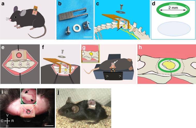
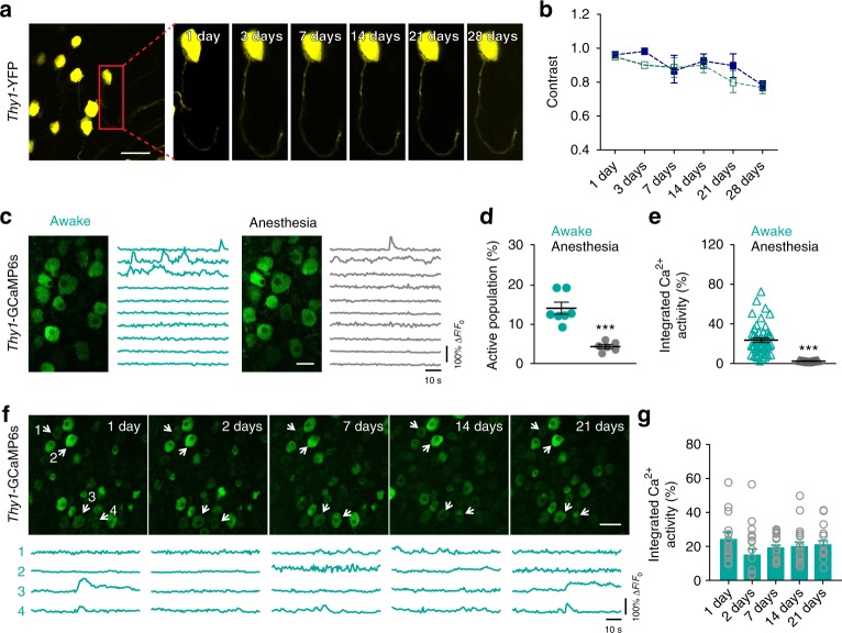
The dorsal root ganglia (DRG) contain the somas of first-order sensory neurons critical for somatosensation. Due to technical difficulties, DRG neuronal activity in awake behaving animals remains unknown. Here, we develop a method for imaging DRG at cellular and subcellular resolution over weeks in awake mice. The method involves the installation of an intervertebral fusion mount to reduce spinal movement, and the implantation of a vertebral glass window without interfering animals' motor and sensory functions. In vivo two-photon calcium imaging shows that DRG neuronal activity is higher in awake than anesthetized animals. Immediately after plantar formalin injection, DRG neuronal activity increases substantially and this activity upsurge correlates with animals' phasic pain behavior. Repeated imaging of DRG over 5 weeks after formalin injection reveals persistent neuronal hyperactivity associated with ongoing pain. The method described here provides an important means for in vivo studies of DRG functions in sensory perception and disorders.
背根神经节(DRG)包含对躯体感觉至关重要的一级感觉神经元的体。由于技术上的困难,清醒行为动物的 DRG 神经元活动仍然未知。在这里,我们开发了一种在清醒小鼠中数周内以细胞和亚细胞分辨率对 DRG 进行成像的方法。该方法涉及安装椎间融合装置以减少脊柱运动,以及植入不会干扰动物运动和感觉功能的椎骨玻璃窗口。体内双光子钙成像显示,清醒动物的 DRG 神经元活动高于麻醉动物。在足底福尔马林注射后立即,DRG 神经元活动显著增加,这种活动增加与动物的阵发性疼痛行为相关。福尔马林注射后 5 周内对 DRG 进行重复成像显示与持续性疼痛相关的持续神经元过度活跃。这里描述的方法为感觉感知和障碍中 DRG 功能的体内研究提供了重要手段。