Orthopaedic Surgery, Osaka University Graduate School of Medicine, Suita, Osaka, Japan.
Anesthesiology and Intensive Care Medicine, Osaka University Graduate School of Medicine, Suita, Osaka, Japan.
Sci Rep. 2019 Jul 18;9(1):10456. doi: 10.1038/s41598-019-46859-5.
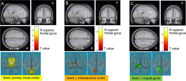
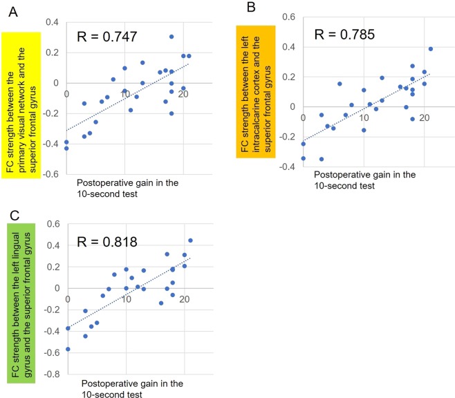
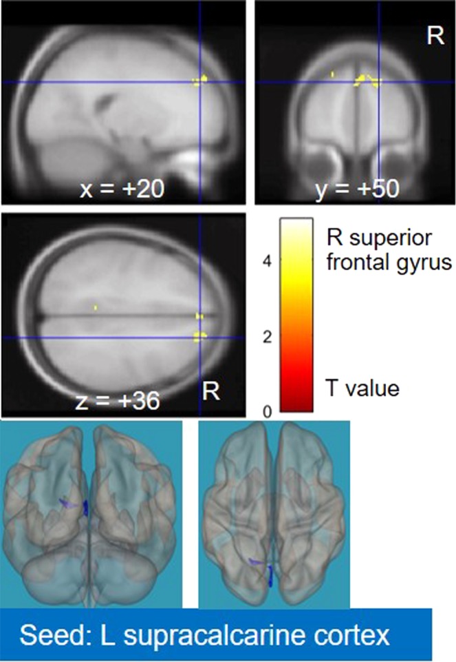
Recently, there has been increasing interest in strategies to predict neurological recovery in cervical myelopathy (CM) based on clinical images of the cervical spine. In this study, we aimed to explore potential preoperative brain biomarkers that can predict postoperative neurological recovery in CM patients by using resting-state functional magnetic resonance imaging (rs-fMRI) and functional connectivity (FC) analysis. Twenty-eight patients with CM and 28 age- and sex-matched healthy controls (HCs) underwent rs-fMRI (twice for CM patients, before and six months after surgery). A seed-to-voxel analysis was performed, and the following three statistical analyses were conducted: (i) FC comparisons between preoperative CM and HC; (ii) correlation analysis between preoperative FCs and clinical scores; and (iii) postoperative FC changes in CM. Our analyses identified three FCs between the visual cortex and the right superior frontal gyrus based on the conjunction of the first two analyses [(i) and (ii)]. These FCs may act as potential biomarkers for postoperative gain in the 10-second test and might be sufficient to provide a prediction formula for potential recovery. Our findings provide preliminary evidence supporting the possibility of novel predictive measures for neurological recovery in CM using rs-fMRI.
最近,人们越来越关注基于颈椎影像学的策略,以预测颈椎病(CM)的神经恢复。在这项研究中,我们旨在通过静息态功能磁共振成像(rs-fMRI)和功能连接(FC)分析,探讨潜在的术前脑生物标志物,以预测 CM 患者术后的神经恢复。28 例 CM 患者和 28 例年龄和性别匹配的健康对照者(HCs)接受了 rs-fMRI(CM 患者两次,手术前和手术后 6 个月)。进行了种子到体素分析,并进行了以下三个统计分析:(i)术前 CM 与 HC 之间的 FC 比较;(ii)术前 FCs 与临床评分的相关性分析;(iii)CM 术后 FC 变化。我们的分析基于前两个分析(i)和(ii),确定了视觉皮层与右侧额上回之间的三个 FC。这些 FC 可能作为术后 10 秒测试增益的潜在生物标志物,足以提供潜在恢复的预测公式。我们的研究结果提供了初步证据,支持使用 rs-fMRI 对 CM 的神经恢复进行新的预测措施的可能性。